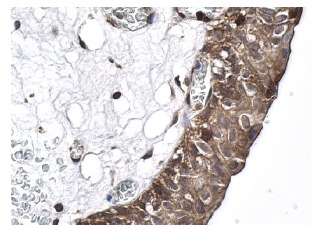
Immunoperoxidase staining of formalin fixed, paraffin-embedded human urinary bladder tissue showing cytoplasmic and nuclear staining of urothelial cells.
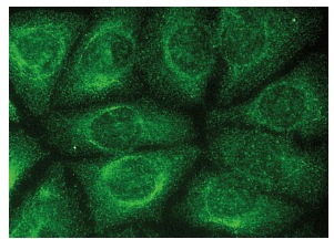
Immunofluorescence staining of methanol-fixed HeLa cells showing cytoplasmic and nuclear localization.
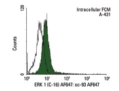
Intracellular FCM analysis of fixed and permeabilized A-431 cells. Black line histogram represents the isotype control, normal rabbit IgG: .
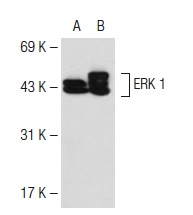
Western blot analysis of ERK 1 expression in non-transfected: (A) and human ERK 1 transfected: (B) 293T whole cell lysates.
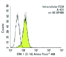
Intracellular FCM analysis of fixed and permeabilized A-431 cells. Black line histogram represents the isotype control, normal rabbit IgG: .
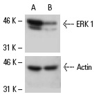
Western blot analysis of ERK 1 expression in non-transfected control (A) and ERK 1 siRNA transfected (B) NIH/3T3 cells. Blot probed with ERK 1 (C-16): . Actin (C-2): used as specificity and loading control.
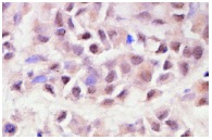
Immunoperoxidase staining of formalin-fixed, paraffin-embedded human breast tumor showing cytoplasmic and nuclear staining.
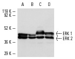
Western blot analysis of ERK 1 and ERK 2 expression in A-431 (A), HeLa (B), KNRK (C) and NIH/3T3 (D) whole cell lysates.
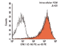
Intracellular FCM analysis of fixed and permeabilized A-431 cells. Black line histogram represents the isotype control, normal rabbit IgG: .
Product Profile
| Product Name | ERK 1 Antibody (C-16) AC |
|---|---|
| Antibody Type | Primary Antibodies |
| Modification Notes | Mitogen-activated protein kinase (MAPK) signaling pathways involve two closely related MAP kinases, known as extracellular-signal-related kinase 1 (ERK 1, p44) and 2 (ERK 2, p42). Growth factors, steroid hormones, G protein-coupled receptor ligands and neurotransmitters can initiate MAPK signaling pathways. Activation of ERK 1 and ERK 2 requires phosphorylation by upstream kinases such as MAP kinasekinase (MEK), MEK kinase and Raf-1. ERK 1 and ERK 2 phosphorylation can occur at specific tyrosine and threonine sites mapping within consensus motifs that include the threonine-glutamate-tyrosine motif. ERK activation leads to dimerization with other ERKs and subsequent localization to the nucleus. Active ERK dimers phosphorylate serine and threonine residues on nuclear proteins and influence a host of responses that include proliferation, differentiation, transcription regulation and development. The human ERK 1 gene maps to chromosome 16p12-p11.2 and encodes a 379 amino acid protein that shares 83% sequence identity to ERK 2. |
Key Feature
| Clonality | Polyclonal |
|---|---|
| Isotype | IgG |
| Host Species | Rabbit |
| Tested Applications | |
recommended for detection of ERK 1 p44 and, to a lesser extent, ERK 2 p42 of mouse, rat, human, avian, frog and zebrafish origin by WB, IP, IF, IHC(P), FCM and ELISA; also reactive with additional species, including equine, canine, bovine and porcine: |
|
| Species Reactivity | |
| Concentration | 1mg/ml |
| Purification | Affinity purified |
Target Information
| Alternative Names | epitope mapping at the C-terminus of ERK 1 of rat origin |
|---|---|
| Tissue Specificity | epitope mapping at the C-terminus of ERK 1 of rat origin |
Database Links
| Entrez Gene | 5595 |
|---|
Application
-

Application
Immunoperoxidase staining of formalin fixed, paraffin-embedded human urinary bladder tissue showing cytoplasmic and nuclear staining of urothelial cells.
-

Application
Immunofluorescence staining of methanol-fixed HeLa cells showing cytoplasmic and nuclear localization.
-

Application
Intracellular FCM analysis of fixed and permeabilized A-431 cells. Black line histogram represents the isotype control, normal rabbit IgG: .
-

Application
Western blot analysis of ERK 1 expression in non-transfected: (A) and human ERK 1 transfected: (B) 293T whole cell lysates.
-

Application
Intracellular FCM analysis of fixed and permeabilized A-431 cells. Black line histogram represents the isotype control, normal rabbit IgG: .
-

Application
Western blot analysis of ERK 1 expression in non-transfected control (A) and ERK 1 siRNA transfected (B) NIH/3T3 cells. Blot probed with ERK 1 (C-16): . Actin (C-2): used as specificity and loading control.
-

Application
Immunoperoxidase staining of formalin-fixed, paraffin-embedded human breast tumor showing cytoplasmic and nuclear staining.
-

Application
Western blot analysis of ERK 1 and ERK 2 expression in A-431 (A), HeLa (B), KNRK (C) and NIH/3T3 (D) whole cell lysates.
-

Application
Intracellular FCM analysis of fixed and permeabilized A-431 cells. Black line histogram represents the isotype control, normal rabbit IgG: .
| Application Notes | recommended for detection of ERK 1 p44 and, to a lesser extent, ERK 2 p42 of mouse, rat, human, avian, frog and zebrafish origin by WB, IP, IF, IHC(P), FCM and ELISA; also reactive with additional species, including equine, canine, bovine and porcine: |
|---|
Additional Information
| Form | Liquid |
|---|---|
| Storage Instructions | For short-term storage, store at 4° C. For long-term storage, aliquot and store at -20ºC or below. Avoid multiple freeze-thaw cycles. |
| Storage Buffer | phosphate buffered saline , pH 7.4, 150mM NaCl, 0.02% sodium azide and 50% glycerol. |
- Related products
- Camel Growth Hormone (GH) ELISA Kit OM642709
- Camel Insulin-like growth factors 1 (IGF-1) ELISA Kit OM642708
- Human HIV-1 p24 core protein (HIV-1 p24) antibody ELISA Kit OM642706
- Antigen Repair 20X pH6.0 OM642703
- 647 labeled Tyramide OM642699
-
- ASSAY KITS
-
- SERUM
2013 © Omnimabs , All Rights Reserved.

