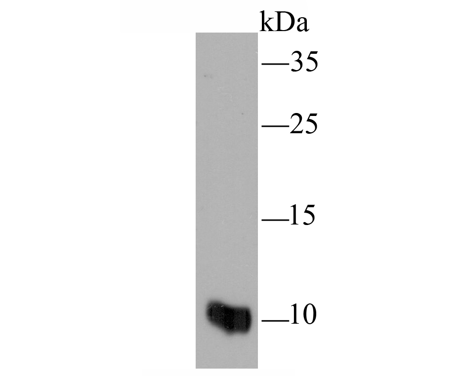
Fig1: Western blot analysis of MRP8/S100A8 on HL-60 cell lysate using anti-MRP8/S100A8 antibody at 1/100 dilution.
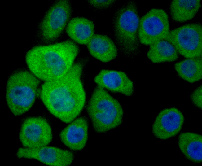
Fig2: ICC staining MRP8/S100A8 in AGS cells (green). The nuclear counter stain is DAPI (blue). Cells were fixed in paraformaldehyde, permeabilised with 0.25% Triton X100/PBS.
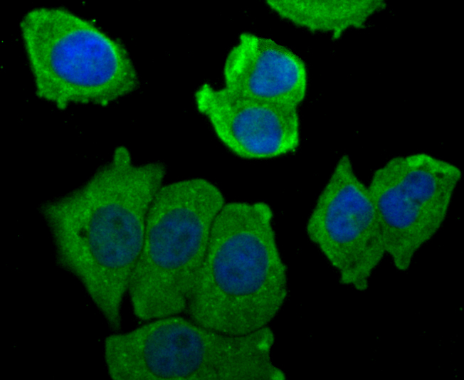
Fig3: ICC staining MRP8/S100A8 in MCF-7 cells (green). The nuclear counter stain is DAPI (blue). Cells were fixed in paraformaldehyde, permeabilised with 0.25% Triton X100/PBS.
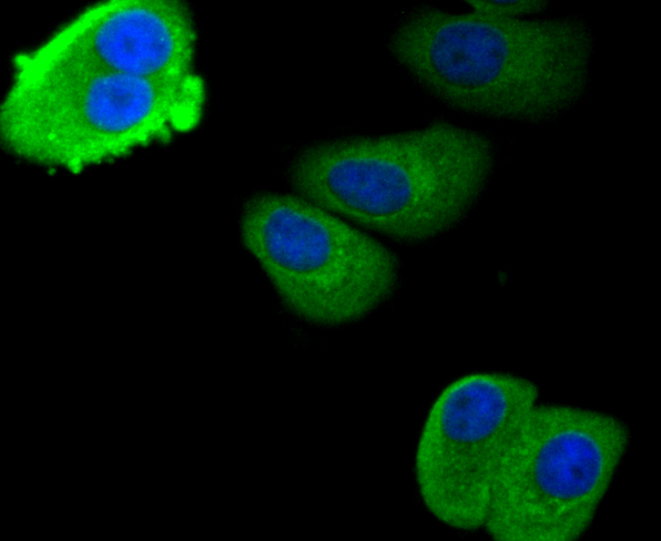
Fig4: ICC staining MRP8/S100A8 in SK-Br-3 cells (green). The nuclear counter stain is DAPI (blue). Cells were fixed in paraformaldehyde, permeabilised with 0.25% Triton X100/PBS.
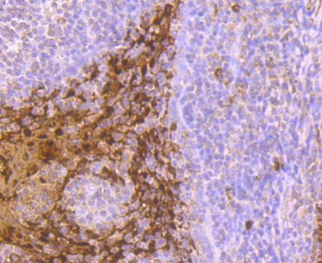
Fig5: Immunohistochemical analysis of paraffin-embedded human tonsil tissue using anti-MRP8/S100A8 antibody. Counter stained with hematoxylin.
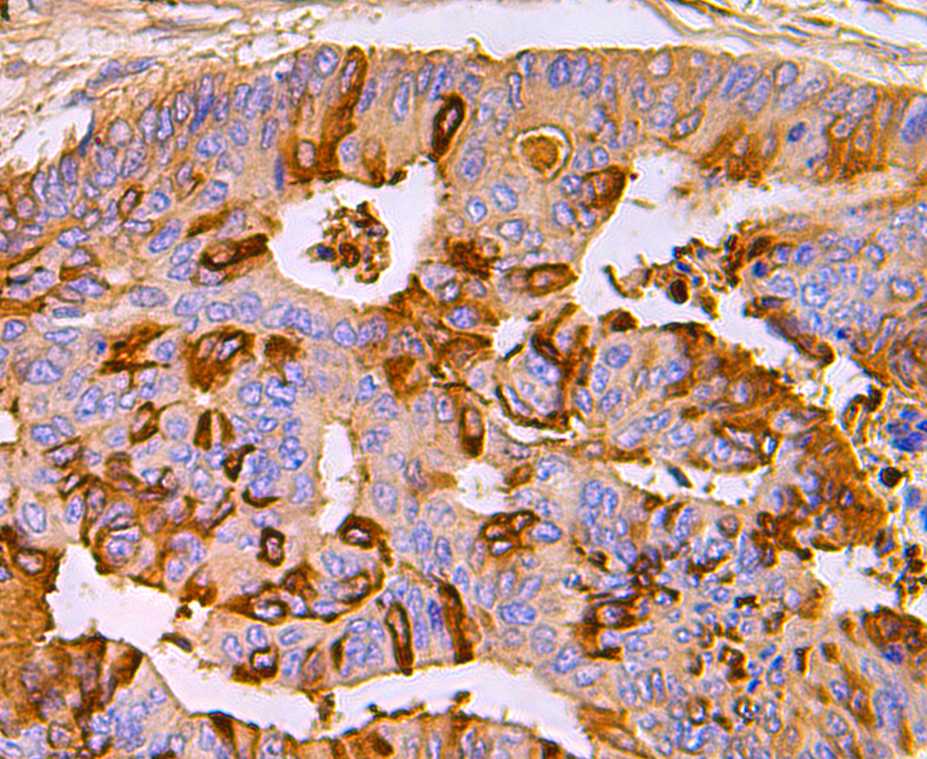
Fig6: Immunohistochemical analysis of paraffin-embedded human colon cancer tissue using anti-MRP8/S100A8 antibody. Counter stained with hematoxylin.
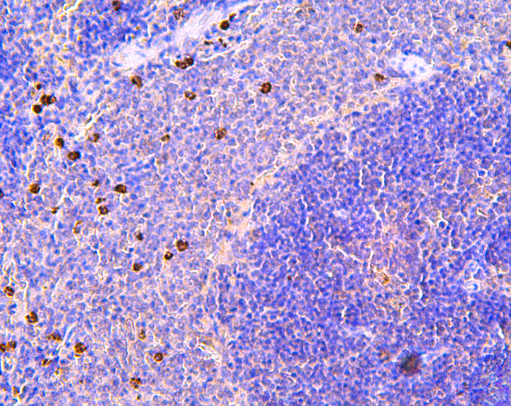
Fig7: Immunohistochemical analysis of paraffin-embedded rat spleen tissue using anti-MRP8/S100A8 antibody. Counter stained with hematoxylin.
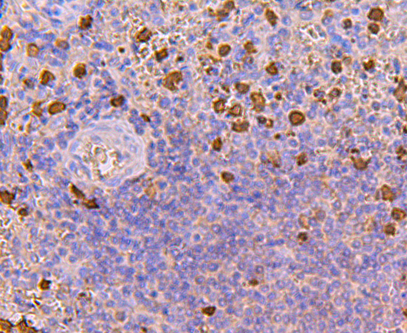
Fig8: Immunohistochemical analysis of paraffin-embedded human spleen tissue using anti-MRP8/S100A8 antibody. Counter stained with hematoxylin.
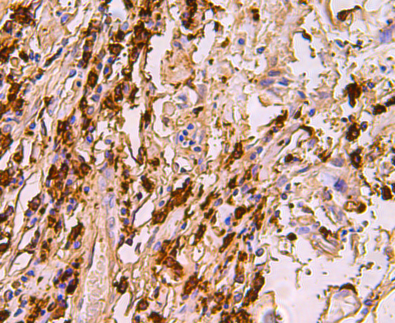
Fig9: Immunohistochemical analysis of paraffin-embedded human breast cancer tissue using anti-MRP8/S100A8 antibody. Counter stained with hematoxylin.
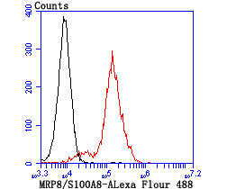
Fig10: Flow cytometric analysis of THP-1 cells with MRP8/S100A8 antibody at 1/100 dilution (red) compared with an unlabelled control (cells without incubation with primary antibody; black).
Product Profile
| Product Name | MRP8/S100A8 |
|---|---|
| Antibody Type | Primary Antibodies |
| Product description |
|
| Immunogen | Recombinant protein |
Key Feature
| Clonality | Polyclonal |
|---|---|
| Isotype | IgG |
| Host Species | Rabbit |
| Tested Applications | |
WB:1:500 ICC:1:50-1:200 IHC:1:50-1:200 FC:1:50-1:100 |
|
| Species Reactivity | |
| Concentration | 1 mg/mL. |
Target Information
| Alternative Names | 60B8Ag antibody
AI323541 antibody
B8Ag antibody
BEE11 antibody
CAGA antibody
Calgranulin-A antibody
Calprotectin L1L subunit antibody
Calprotectin included antibody CFAG antibody CGLA antibody Chemotactic cytokine CP-10 antibody CP-10 antibody Cystic fibrosis antigen antibody L1Ag antibody Leukocyte L1 complex light chain antibody MA387 antibody MIF antibody Migration inhibitory factor-related protein 8 antibody MRP-8 antibody Myeloid-related protein 8 antibody Neutrophil cytosolic 7 kDa protein antibody NIF antibody p8 antibody Pro-inflammatory S100 cytokine antibody Protein S100-A8 antibody S100 calcium binding protein A8 (calgranulin A) antibody S100 calcium binding protein A8 antibody S100 calcium-binding protein A8 antibody S100A8 antibody S100A8/S100A9 complex included antibody S10A8_HUMAN antibody Urinary stone protein band A antibody |
|---|---|
| Molecular Weight(MW) | 11 kDa |
| Cellular Localization | Cell membrane. Cytoplasm. Secreted. |
Database Links
| SwissProt ID | P05109 P27005 |
|---|
Application
-

Application
Fig1: Western blot analysis of MRP8/S100A8 on HL-60 cell lysate using anti-MRP8/S100A8 antibody at 1/100 dilution.
-

Application
Fig2: ICC staining MRP8/S100A8 in AGS cells (green). The nuclear counter stain is DAPI (blue). Cells were fixed in paraformaldehyde, permeabilised with 0.25% Triton X100/PBS.
-

Application
Fig3: ICC staining MRP8/S100A8 in MCF-7 cells (green). The nuclear counter stain is DAPI (blue). Cells were fixed in paraformaldehyde, permeabilised with 0.25% Triton X100/PBS.
-

Application
Fig4: ICC staining MRP8/S100A8 in SK-Br-3 cells (green). The nuclear counter stain is DAPI (blue). Cells were fixed in paraformaldehyde, permeabilised with 0.25% Triton X100/PBS.
-

Application
Fig5: Immunohistochemical analysis of paraffin-embedded human tonsil tissue using anti-MRP8/S100A8 antibody. Counter stained with hematoxylin.
-

Application
Fig6: Immunohistochemical analysis of paraffin-embedded human colon cancer tissue using anti-MRP8/S100A8 antibody. Counter stained with hematoxylin.
-

Application
Fig7: Immunohistochemical analysis of paraffin-embedded rat spleen tissue using anti-MRP8/S100A8 antibody. Counter stained with hematoxylin.
-

Application
Fig8: Immunohistochemical analysis of paraffin-embedded human spleen tissue using anti-MRP8/S100A8 antibody. Counter stained with hematoxylin.
-

Application
Fig9: Immunohistochemical analysis of paraffin-embedded human breast cancer tissue using anti-MRP8/S100A8 antibody. Counter stained with hematoxylin.
-

Application
Fig10: Flow cytometric analysis of THP-1 cells with MRP8/S100A8 antibody at 1/100 dilution (red) compared with an unlabelled control (cells without incubation with primary antibody; black).
| Positive Control | HL-60, AGS, MCF-7, SK-Br-3, human tonsil tissue, human colon cancer tissue, human spleen tissue, human breast cancer tissue, rat spleen tissue, THP-1. |
|---|---|
| Application Notes | WB:1:500 ICC:1:50-1:200 IHC:1:50-1:200 FC:1:50-1:100 |
Additional Information
| Form | Liquid |
|---|---|
| Storage Instructions | Store at +4℃ after thawing. Aliquot store at -20℃ or -80℃. Avoid repeated freeze / thaw cycles. |
| Storage Buffer | 1*TBS (pH7.4), 0.5%BSA, 50%Glycerol. Preservative: 0.05% Sodium Azide. |
- Related products
- Camel Growth Hormone (GH) ELISA Kit OM642709
- Camel Insulin-like growth factors 1 (IGF-1) ELISA Kit OM642708
- Human HIV-1 p24 core protein (HIV-1 p24) antibody ELISA Kit OM642706
- Antigen Repair 20X pH6.0 OM642703
- 647 labeled Tyramide OM642699
-
- ASSAY KITS
-
- SERUM
2013 © Omnimabs , All Rights Reserved.

