Immunohistochemical
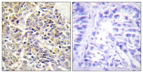
Immunohistochemistry analysis of paraffin-embedded human lung carcinoma tissue, using ASC Antibody. The picture on the right is treated with the synthesized peptide.Western blot
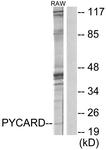
Western blot analysis of extracts from RAW264.7 cells, using ASC Antibody. The lane on the right is treated with the synthesized peptideWB
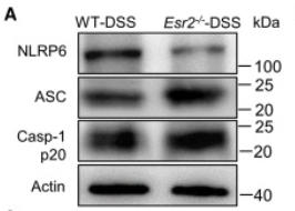
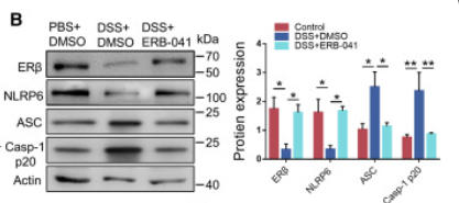
(A) NLRP6, ASC, and Casp-1 p20 expression of mouse colon homogenate.(I) ERβ, NLRP6, ASC, and Casp-1 p20 expression of mice colon homogenate. See also Figure S1 and Data S1.WB
(B) Immunoblotting of treated cell lysates. Data are representative of three independent experiments. ∗p < 0.05, ∗∗p < 0.01, by two-way ANOVA with Tukey’s post hoc analysis. Normal NCM-460 cells were stimulated with the selective ERβ agonist (ERB-041, 1 μM) and ERα agonist (PPT, 1 μM) for 48 h.WB
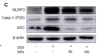
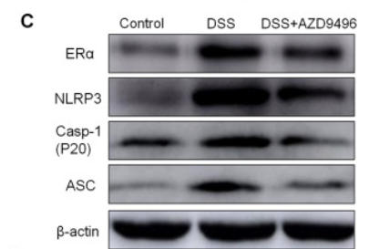
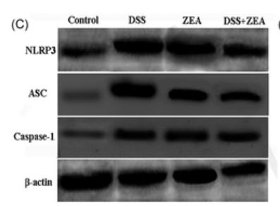
(C) Protein expression of NLRP3, ASC, Caspase-1 and ERα in colon tissues.WB
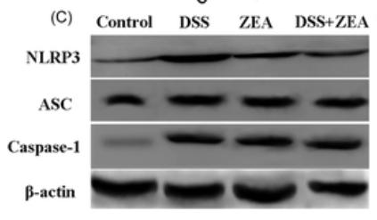
(C) Protein levels of NLRP3, ASC and Caspase-1 in colon tissues.WB
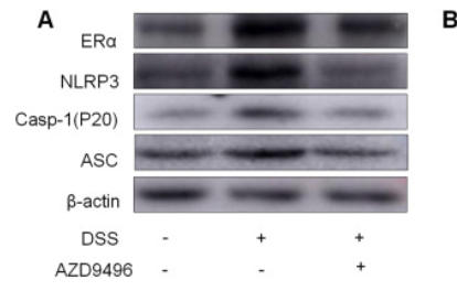
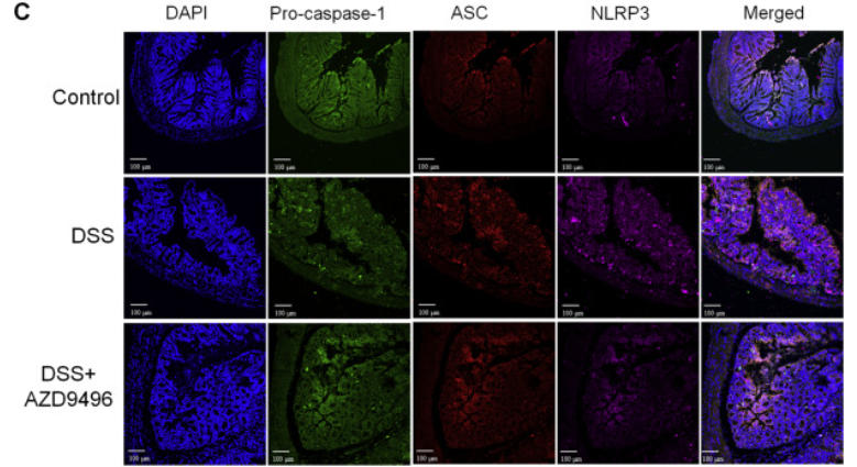
(A) Protein levels of NLRP3, ASC and Caspase-1 in colon tissues.(C) Colon tissue was immunofluorescently stained with Cy5-NLRP3 antibody, PE-ASC antibody and FITC-pro-Caspase-1 antibody.WB
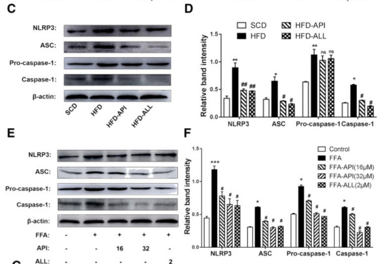
(C-D) NLRP3, ASC, pro-caspase-1, caspase-1 protein levels were detected by Western blots and quantified by using Image J software in mice. The data are expressed as the mean±S.E.M. (n=6). *P<.05, **P<.01, ***P<.01 vs. The SCD group; #P<.05 ##P<.01 vs. the HFD group. (E-F) NLRP3, ASC, Pro-caspase-1, caspase-1 protein levels were detected by Western blots and quantified by using Image J software in hepa1–6 cells.WB
C, Protein expression levels of NLRP3, ASC, and caspase-1 in mouse colon tissue.WB
C, NLRP3, ASC, and caspase-1 protein expression in IPEC-J2 cells.IF
(C) Colon tissue was immunofluorescently stained with Cy5-NLRP3 antibody, PE-ASC antibody and FITC-pro-Caspase-1 antibody.| Product Name | Rabbit anti-ASC polyclonal antibody |
|---|---|
| Antibody Type | Primary Antibodies |
| Immunogen | The antiserum was produced against synthesized peptide derived from human ASC. |
| Clonality | Polyclonal |
|---|---|
| Host Species | Rabbit |
| Tested Applications | ELISAIHCWB |
| WB:1:500~1:1000 IHC:1:50~1:100 ELISA:1:40000 |
|
| Species Reactivity | HumanMouseRat |
| Concentration | 1mg/ml |
| Purification | Affinity purified |
| Gene Symbol | PYCARD |
|---|---|
| Gene Synonyms | ASC ASC apoptosis-associated speck-like protein containing a CARD CARD5 caspase recruitment domain protein 5 TMS1 target of methylation-induced silencing 1 |
Immunohistochemical
Immunohistochemistry analysis of paraffin-embedded human lung carcinoma tissue, using ASC Antibody. The picture on the right is treated with the synthesized peptide.
Western blot
Western blot analysis of extracts from RAW264.7 cells, using ASC Antibody. The lane on the right is treated with the synthesized peptide
WB
(A) NLRP6, ASC, and Casp-1 p20 expression of mouse colon homogenate.(I) ERβ, NLRP6, ASC, and Casp-1 p20 expression of mice colon homogenate. See also Figure S1 and Data S1.
WB
(B) Immunoblotting of treated cell lysates. Data are representative of three independent experiments. ∗p < 0.05, ∗∗p < 0.01, by two-way ANOVA with Tukey’s post hoc analysis. Normal NCM-460 cells were stimulated with the selective ERβ agonist (ERB-041, 1 μM) and ERα agonist (PPT, 1 μM) for 48 h.
WB
(C) Protein expression of NLRP3, ASC, Caspase-1 and ERα in colon tissues.
WB
(C) Protein levels of NLRP3, ASC and Caspase-1 in colon tissues.
WB
(A) Protein levels of NLRP3, ASC and Caspase-1 in colon tissues.(C) Colon tissue was immunofluorescently stained with Cy5-NLRP3 antibody, PE-ASC antibody and FITC-pro-Caspase-1 antibody.
WB
(C-D) NLRP3, ASC, pro-caspase-1, caspase-1 protein levels were detected by Western blots and quantified by using Image J software in mice. The data are expressed as the mean±S.E.M. (n=6). *P<.05, **P<.01, ***P<.01 vs. The SCD group; #P<.05 ##P<.01 vs. the HFD group. (E-F) NLRP3, ASC, Pro-caspase-1, caspase-1 protein levels were detected by Western blots and quantified by using Image J software in hepa1–6 cells.
WB
C, Protein expression levels of NLRP3, ASC, and caspase-1 in mouse colon tissue.
WB
C, NLRP3, ASC, and caspase-1 protein expression in IPEC-J2 cells.
IF
(C) Colon tissue was immunofluorescently stained with Cy5-NLRP3 antibody, PE-ASC antibody and FITC-pro-Caspase-1 antibody.| Application Notes | WB:1:500~1:1000 IHC:1:50~1:100 ELISA:1:40000 |
|---|
| Form | Liquid |
|---|---|
| Storage Instructions | store at -20°C. Avoid repeated freeze / thaw cycles |
| Storage Buffer | Rabbit IgG in phosphate buffered saline (without Mg2+ and Ca2+), pH 7.4, 150mM NaCl, 0.02% sodium azide and 50% glycerol. |


