Application
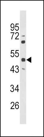
Western blot analysis of PAX6 Antibody (Center) in Hela cell line lysates (35ug/lane). PAX6 (arrow) was detected using the purified Pab.Application
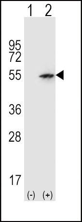
Western blot analysis of PAX6 (arrow) using rabbit polyclonal PAX6 Antibody (Center) . 293 cell lysates (2 ug/lane) either nontransfected (Lane 1) or transiently transfected (Lane 2) with the PAX6 gene.Application
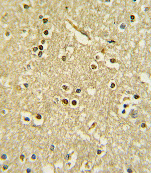
Formalin-fixed and paraffin-embedded human brain tissue with PAX6 Antibody (Center), which was peroxidase-conjugated to the secondary antibody, followed by DAB staining. This data demonstrates the use of this antibody for immunohistochemistry; clinical relevance has not been evaluated.Application
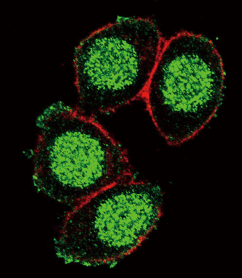
Confocal immunofluorescent analysis of PAX6 Antibody (Center)(Cat#AP6929c) with Hela cell followed by Alexa Fluor 488-conjugated goat anti-rabbit lgG (green). Actin filaments have been labeled with Alexa Fluor 555 phalloidin (red).Application
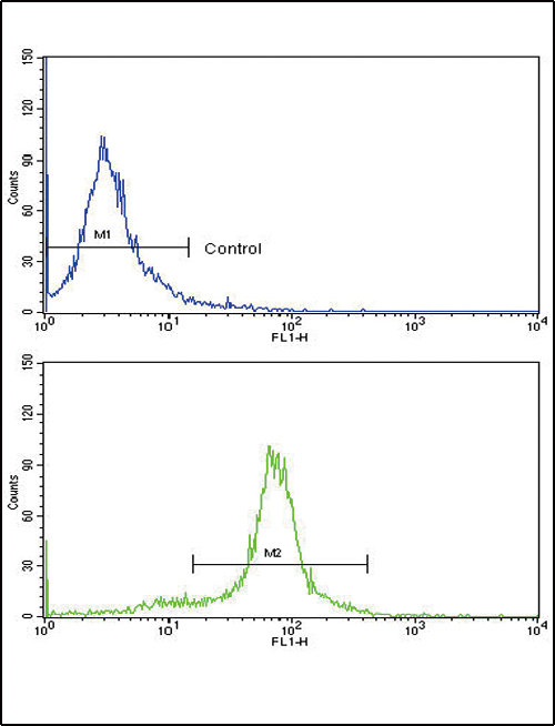
Flow cytometric analysis of hela cells using PAX6 Antibody (Center)(bottom histogram) compared to a negative control cell (top histogram). FITC-conjugated goat-anti-rabbit secondary antibodies were used for the analysis.| Product Name | PAX6 Antibody (Center) |
|---|---|
| Antibody Type | Primary Antibodies |
| Antigen Alias | PAX6; AN2; Paired box protein Pax-6; Aniridia type II protein; Oculorhombin |
| Clonality | Polyclonal |
|---|---|
| Isotype | Ig |
| Host Species | Rabbit |
| Tested Applications | WBIHCIFFC |
| WB:1:100~500 IHC |
|
| Species Reactivity | Human |
| Concentration | 1mg/ml |
| Gene Synonyms | AN2 |
|---|---|
| Alternative Names | PAX6 AN2 Paired box protein Pax-6 Aniridia type II protein Oculorhombin |
| Molecular Weight(MW) | 46683 Da |
| Function | Transcription factor with important functions in the development of the eye, nose, central nervous system and pancreas Required for the differentiation of pancreatic islet alpha cells (By similarity). Competes with PAX4 in binding to a common element in the glucagon, insulin and somatostatin promoters. Regulates specification of the ventral neuron subtypes by establishing the correct progenitor domains (By similarity). Isoform 5a appears to function as a molecular switch that specifies target genes |
| Tissue Specificity | Fetal eye, brain, spinal cord and olfactory epithelium. Isoform 5a is less abundant than the PAX6 shorter form |
| Cellular Localization | Nucleus. |
| Entrez Gene | 5080 |
|---|
Application
Western blot analysis of PAX6 Antibody (Center) in Hela cell line lysates (35ug/lane). PAX6 (arrow) was detected using the purified Pab.
Application
Western blot analysis of PAX6 (arrow) using rabbit polyclonal PAX6 Antibody (Center) . 293 cell lysates (2 ug/lane) either nontransfected (Lane 1) or transiently transfected (Lane 2) with the PAX6 gene.
Application
Formalin-fixed and paraffin-embedded human brain tissue with PAX6 Antibody (Center), which was peroxidase-conjugated to the secondary antibody, followed by DAB staining. This data demonstrates the use of this antibody for immunohistochemistry; clinical relevance has not been evaluated.
Application
Confocal immunofluorescent analysis of PAX6 Antibody (Center)(Cat#AP6929c) with Hela cell followed by Alexa Fluor 488-conjugated goat anti-rabbit lgG (green). Actin filaments have been labeled with Alexa Fluor 555 phalloidin (red).
Application
Flow cytometric analysis of hela cells using PAX6 Antibody (Center)(bottom histogram) compared to a negative control cell (top histogram). FITC-conjugated goat-anti-rabbit secondary antibodies were used for the analysis.| Application Notes | WB:1:100~500 IHC |
|---|
| Form | Liquid |
|---|---|
| Storage Instructions | For short-term storage, store at 4° C. For long-term storage, aliquot and store at -20ºC or below. Avoid multiple freeze-thaw cycles. |
| Storage Buffer | Purified polyclonal antibody supplied in PBS with 0.09% (W/V) sodium azide. This antibody is purified through a protein A column, followed by peptide affinity purification. |


