Application
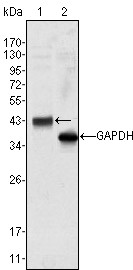
Figure 1: Western blot analysis using anti-WNT10B mAb against Hela cell lysate (1).Application
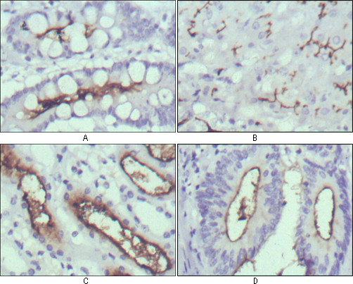
Figure 2: Immunohistochemical analysis of paraffin-embedded human normal stomach (A), normal liver (B), normal kidney (C) and rectum cancer tissues (D) using anti-WNT10B mAb with DAB staining.Application
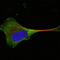
Figure 3: Confocal immunofluorescence analysis of PANC-1 cells using anti-WNT10B mAb (green). Red: Actin filaments have been labeled with Alexa Fluor-555 phalloidin. Blue: DRAQ5 fluorescent DNA dye.| Product Name | WNT10B Antibody |
|---|---|
| Antibody Type | Primary Antibodies |
| Antigen Alias | SHFM6; WNT-12 |
| Clonality | Monoclonal |
|---|---|
| Isotype | IgG1 |
| Clone Number | 5A7 |
| Host Species | Mouse |
| Tested Applications | WBIHCIF |
| WB:1:500 - 2000. IHC |
|
| Species Reactivity | Human |
| Concentration | 1mg/ml |
| Gene Synonyms | WNT12 |
|---|---|
| Alternative Names | SHFM6 WNT-12 |
| Molecular Weight(MW) | 43000 Da |
| Function | Ligand for members of the frizzled family of seven transmembrane receptors. Probable developmental protein. May be a signaling molecule which affects the development of discrete regions of tissues. Is likely to signal over only few cell diameters (By similarity) |
| Tissue Specificity | Detected in most adult tissues. Highest levels were found in heart and skeletal muscle. Low levels are found in brain |
| Cellular Localization | Secreted, extracellular space, extracellular matrix |
| Entrez Gene | 7480 |
|---|---|
| Nucleotide Accession | NP_003385.2 |
Application
Figure 1: Western blot analysis using anti-WNT10B mAb against Hela cell lysate (1).
Application
Figure 2: Immunohistochemical analysis of paraffin-embedded human normal stomach (A), normal liver (B), normal kidney (C) and rectum cancer tissues (D) using anti-WNT10B mAb with DAB staining.
Application
Figure 3: Confocal immunofluorescence analysis of PANC-1 cells using anti-WNT10B mAb (green). Red: Actin filaments have been labeled with Alexa Fluor-555 phalloidin. Blue: DRAQ5 fluorescent DNA dye.| Application Notes | WB:1:500 - 2000. IHC |
|---|
| Form | Liquid |
|---|---|
| Storage Instructions | For short-term storage, store at 4° C. For long-term storage, aliquot and store at -20ºC or below. Avoid multiple freeze-thaw cycles. |
| Storage Buffer | Purified recombinant fragment of human WNT10B expressed in E. Coli. |


