Application
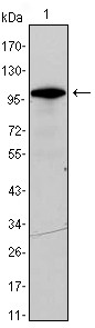
Figure 1: Western blot analysis using DDX4 mAb against DDX4-hIgGFc transfected HEK293 cell lysate.Application
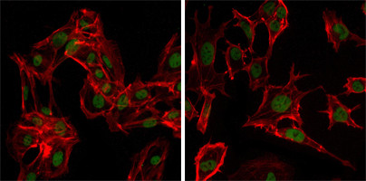
Figure 2:Immunofluorescence analysis of MSCs(left) and NTERA-2 (right) cells using anti-DDX4 mAb (green). Red: Actin filaments have been labeled with DY-554 phalloidin.Application
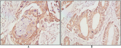
Figure 3: Immunohistochemical analysis of paraffin-embedded human lung cancer (A) and rectal cancer (B), showing cytoplasmic localization using anti-DDX4 antibody with DAB staining.| Product Name | DDX4 Antibody |
|---|---|
| Antibody Type | Primary Antibodies |
| Antigen Alias | VASA;MGC111074;DDX4 |
| Clonality | Monoclonal |
|---|---|
| Isotype | IgG1 |
| Clone Number | 2F9H5 |
| Host Species | Mouse |
| Tested Applications | WBIFIHC |
| WB:1:500 - 2000. IF |
|
| Species Reactivity | Human |
| Concentration | 1mg/ml |
| Gene Synonyms | VASA |
|---|---|
| Alternative Names | VASA MGC111074 DDX4 |
| Molecular Weight(MW) | 79308 Da |
| Function | May play a role in germ cell development. May play a role in sperm motility |
| Tissue Specificity | Expressed only in ovary and testis. Expressed in migratory primordial germ cells in the region of the gonadal ridge in both sexes |
| Cellular Localization | Cytoplasm. Cytoplasm, perinuclear region (By similarity) |
| Entrez Gene | 54514 |
|---|---|
| Nucleotide Accession | NP_077726.1 |
Application
Figure 1: Western blot analysis using DDX4 mAb against DDX4-hIgGFc transfected HEK293 cell lysate.
Application
Figure 2:Immunofluorescence analysis of MSCs(left) and NTERA-2 (right) cells using anti-DDX4 mAb (green). Red: Actin filaments have been labeled with DY-554 phalloidin.
Application
Figure 3: Immunohistochemical analysis of paraffin-embedded human lung cancer (A) and rectal cancer (B), showing cytoplasmic localization using anti-DDX4 antibody with DAB staining.| Application Notes | WB:1:500 - 2000. IF |
|---|
| Form | Liquid |
|---|---|
| Storage Instructions | For short-term storage, store at 4° C. For long-term storage, aliquot and store at -20ºC or below. Avoid multiple freeze-thaw cycles. |
| Storage Buffer | Purified recombinant fragment of human DDX4 expressed in E. Coli. |


