Application
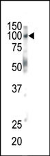
Western blot analysis of anti-EphB1 Pab in mouse brain tissue. EphB1 (arrow) was detected using purified Pab. Secondary HRP-anti-rabbit was used for signal visualization with chemiluminescence.Application
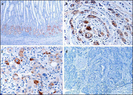
Immunohistochemical analysis of EphB1 in gastric cancer tissues. a EphB1 protein expressed in normal mucosa at the glandular compartment and in a decreasing gradient from the glandular compartment to the foveolar compartment. b EphB1 protein focally positively stained in well-differentiated gastric cancer cells. c EphB1 protein is focally positive in poorly differentiated gastric cancer cells. d Loss of EphB1 expression in gastric cancer cells.(Provided by Jian-dong Wang,Department of Pathology Nanjing Jinling Hospital/Nanjing University School of Medicine)Application
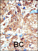
Formalin-fixed and paraffin-embedded human cancer tissue reacted with the primary antibody, which was peroxidase-conjugated to the secondary antibody, followed by DAB staining. This data demonstrates the use of this antibody for immunohistochemistry; clinical relevance has not been evaluated. BC = breast carcinoma; HC = hepatocarcinoma.Application
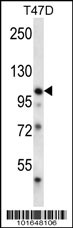
EPHB1 Antibody (H970) western blot analysis in T47D cell line lysates (35ug/lane).This demonstrates the EPHB1 antibody detected the EPHB1 protein (arrow).| Product Name | EphB1 Antibody (C-term) |
|---|---|
| Antibody Type | Primary Antibodies |
| Clonality | Polyclonal |
|---|---|
| Isotype | Ig |
| Host Species | Rabbit |
| Tested Applications | WBIHC |
| WB:1:100~500 IHC |
|
| Species Reactivity | Human |
| Concentration | 1mg/ml |
| Gene Synonyms | ELK, EPHT2, HEK6, NET |
|---|---|
| Alternative Names | EPHB1 ELK EPHT2 HEK6 NET Ephrin type-B receptor 1 EPH tyrosine kinase 2 EPH-like kinase 6 Neuronally-expressed EPH-related tyrosine kinase Tyrosine-protein kinase receptor EPH-2 |
| Molecular Weight(MW) | 109885 Da |
| Function | Receptor tyrosine kinase which binds promiscuously transmembrane ephrin-B family ligands residing on adjacent cells, leading to contact-dependent bidirectional signaling into neighboring cells. The signaling pathway downstream of the receptor is referred to as forward signaling while the signaling pathway downstream of the ephrin ligand is referred to as reverse signaling. Cognate/functional ephrin ligands for this receptor include EFNB1, EFNB2 and EFNB3. During nervous system development, regulates retinal axon guidance redirecting ipsilaterally ventrotemporal retinal ganglion cells axons at the optic chiasm midline. This probably requires repulsive interaction with EFNB2 In the adult nervous system together with EFNB3, regulates chemotaxis, proliferation and polarity of the hippocampus neural progenitors. Beside its role in axon guidance plays also an important redundant role with other ephrin-B receptors in development and maturation of dendritic spines and synapse formation. May also regulate angiogenesis. More generally, may play a role in targeted cell migration and adhesion. Upon activation by EFNB1 and probably other ephrin-B ligands activates the MAPK/ERK and the JNK signaling cascades to regulate cell migration and adhesion respectively |
| Tissue Specificity | Preferentially expressed in brain. |
| Cellular Localization | Cell membrane; Single-pass type I membrane protein. Early endosome membrane. Cell projection, dendrite (By similarity) |
| Entrez Gene | 2047 |
|---|
Application
Western blot analysis of anti-EphB1 Pab in mouse brain tissue. EphB1 (arrow) was detected using purified Pab. Secondary HRP-anti-rabbit was used for signal visualization with chemiluminescence.
Application
Immunohistochemical analysis of EphB1 in gastric cancer tissues. a EphB1 protein expressed in normal mucosa at the glandular compartment and in a decreasing gradient from the glandular compartment to the foveolar compartment. b EphB1 protein focally positively stained in well-differentiated gastric cancer cells. c EphB1 protein is focally positive in poorly differentiated gastric cancer cells. d Loss of EphB1 expression in gastric cancer cells.(Provided by Jian-dong Wang,Department of Pathology Nanjing Jinling Hospital/Nanjing University School of Medicine)
Application
Formalin-fixed and paraffin-embedded human cancer tissue reacted with the primary antibody, which was peroxidase-conjugated to the secondary antibody, followed by DAB staining. This data demonstrates the use of this antibody for immunohistochemistry; clinical relevance has not been evaluated. BC = breast carcinoma; HC = hepatocarcinoma.
Application
EPHB1 Antibody (H970) western blot analysis in T47D cell line lysates (35ug/lane).This demonstrates the EPHB1 antibody detected the EPHB1 protein (arrow).| Application Notes | WB:1:100~500 IHC |
|---|
| Form | Liquid |
|---|---|
| Storage Instructions | For short-term storage, store at 4° C. For long-term storage, aliquot and store at -20ºC or below. Avoid multiple freeze-thaw cycles. |
| Storage Buffer | Purified polyclonal antibody supplied in PBS with 0.09% (W/V) sodium azide. This antibody is purified through a protein G column, eluted with high and low pH buffers and neutralized immediately, followed by dialysis against PBS. |


