Application
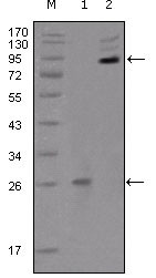
Figure 1: Western blot analysis using anti-EphB2 monoclonal antibody against truncated EphB2 recombinant protein(1) and extracellular EphB2(aa19-476)-hIgGFc transfected CHO-K1 cell lysate(2).Application
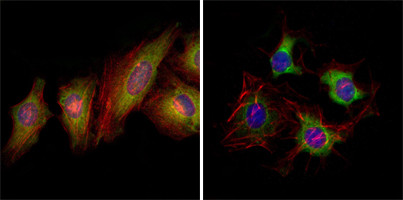
Figure 2:Immunofluorescence analysis of Hale (left) and HepG2 (right) cells using anti-EphB2 mAb (green). Red: Actin filaments have been labeled with DY-554 phalloidin. Blue: DRAQ5 fluorescent DNA dye.| Product Name | EphB2 Antibody |
|---|---|
| Antibody Type | Primary Antibodies |
| Clonality | Monoclonal |
|---|---|
| Isotype | IgG2b |
| Clone Number | 2D12C6 |
| Host Species | Mouse |
| Tested Applications | WBIF |
| WB:1:500 - 2000. IF |
|
| Species Reactivity | Human |
| Concentration | 1mg/ml |
| Gene Synonyms | DRT, EPHT3, EPTH3, ERK, HEK5, TYRO5 |
|---|---|
| Alternative Names | DRT ERK CAPB Hek5 |
| Molecular Weight(MW) | 117493 Da |
| Function | Receptor tyrosine kinase which binds promiscuously transmembrane ephrin-B family ligands residing on adjacent cells, leading to contact-dependent bidirectional signaling into neighboring cells. The signaling pathway downstream of the receptor is referred to as forward signaling while the signaling pathway downstream of the ephrin ligand is referred to as reverse signaling. Functions in axon guidance during development. Involved in the guidance of commissural axons, that form a major interhemispheric connection between the 2 temporal lobes of the cerebral cortex. Also involved in guidance of contralateral inner ear efferent growth cones at the midline and of retinal ganglion cell axons to the optic disk. Beside axon guidance, also regulates dendritic spines development and maturation and stimulates the formation of excitatory synapses. Upon activation by EFNB1, abolishes the ARHGEF15-mediated negative regulation on excitatory synapse formation. Controls other aspects of development including angiogenesis, palate development and in inner ear development through regulation of endolymph production. Forward and reverse signaling through the EFNB2/EPHB2 complex regulate movement and adhesion of cells that tubularize the urethra and septate the cloaca. May function as a tumor suppressor |
| Tissue Specificity | Brain, heart, lung, kidney, placenta, pancreas, liver and skeletal muscle. Preferentially expressed in fetal brain |
| Cellular Localization | Cell membrane; Single-pass type I membrane protein. Cell projection, axon (By similarity). Cell projection, dendrite (By similarity) |
| Entrez Gene | 2048 |
|---|---|
| Nucleotide Accession | NP_059145.2 |
Application
Figure 1: Western blot analysis using anti-EphB2 monoclonal antibody against truncated EphB2 recombinant protein(1) and extracellular EphB2(aa19-476)-hIgGFc transfected CHO-K1 cell lysate(2).
Application
Figure 2:Immunofluorescence analysis of Hale (left) and HepG2 (right) cells using anti-EphB2 mAb (green). Red: Actin filaments have been labeled with DY-554 phalloidin. Blue: DRAQ5 fluorescent DNA dye.| Application Notes | WB:1:500 - 2000. IF |
|---|
| Form | Liquid |
|---|---|
| Storage Instructions | For short-term storage, store at 4° C. For long-term storage, aliquot and store at -20ºC or below. Avoid multiple freeze-thaw cycles. |
| Storage Buffer | Purified recombinant fragment of EphB2(aa17-200) expressed in E. Coli. |


