Application
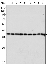
Figure 1: Western blot analysis using anti-GAPDH mAb against Hela (1), A549 (2), A431 (3), MCF-7 (4), K562 (5), Jurkat (6), HL60 (7), SKN-SH (8) and SKBR-3 (9) cell lysate.Application
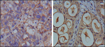
Figure 2: Immunohistochemical analysis of paraffin-embedded human breast carcinoma (left) and kidney carcinoma (right), showing cytoplasmic localization using anti-GAPDH mAb with DAB staining.Application
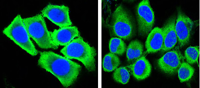
Figure 3: Confocal immunofluorescence analysis of methanol-fixed HepG2 (left) and Hela (right) cells using anti-GAPDH mAb (green), showing cytoplasmic localization. Blue: DRAQ5 fluorescent DNA dye.| Product Name | GAPDH Antibody |
|---|---|
| Antibody Type | Primary Antibodies |
| Clonality | Monoclonal |
|---|---|
| Isotype | IgG1 |
| Clone Number | 1A10 |
| Host Species | Mouse |
| Tested Applications | WBIHCIF |
| WB:1:500 - 2000. IHC |
|
| Species Reactivity | Human |
| Concentration | 1mg/ml |
| Gene Synonyms | GAPD |
|---|---|
| Alternative Names | G3PD GAPD MGC88685 |
| Molecular Weight(MW) | 36053 Da |
| Function | Has both glyceraldehyde-3-phosphate dehydrogenase and nitrosylase activities, thereby playing a role in glycolysis and nuclear functions, respectively. Participates in nuclear events including transcription, RNA transport, DNA replication and apoptosis. Nuclear functions are probably due to the nitrosylase activity that mediates cysteine S-nitrosylation of nuclear target proteins such as SIRT1, HDAC2 and PRKDC. Modulates the organization and assembly of the cytoskeleton. Facilitates the CHP1-dependent microtubule and membrane associations through its ability to stimulate the binding of CHP1 to microtubules (By similarity). Glyceraldehyde-3-phosphate dehydrogenase is a key enzyme in glycolysis that catalyzes the first step of the pathway by converting D-glyceraldehyde 3-phosphate (G3P) into 3-phospho-D- glyceroyl phosphate. Component of the GAIT (gamma interferon- activated inhibitor of translation) complex which mediates interferon-gamma-induced transcript-selective translation inhibition in inflammation processes. Upon interferon-gamma treatment assembles into the GAIT complex which binds to stem loop-containing GAIT elements in the 3'-UTR of diverse inflammatory mRNAs (such as ceruplasmin) and suppresses their translation |
| Tissue Specificity | Purified recombinant fragment of human GAPDH expressed in E. Coli. |
| Cellular Localization | Cytoplasm, cytosol. Nucleus (By similarity). Cytoplasm, perinuclear region. Membrane. Cytoplasm, cytoskeleton (By similarity). Note=Translocates to the nucleus following S- nitrosylation and interaction with SIAH1, which contains a nuclear localization signal (By similarity). Postnuclear and Perinuclear regions |
| Entrez Gene | 2597 |
|---|---|
| Nucleotide Accession | NP_002037.2 |
Application
Figure 1: Western blot analysis using anti-GAPDH mAb against Hela (1), A549 (2), A431 (3), MCF-7 (4), K562 (5), Jurkat (6), HL60 (7), SKN-SH (8) and SKBR-3 (9) cell lysate.
Application
Figure 2: Immunohistochemical analysis of paraffin-embedded human breast carcinoma (left) and kidney carcinoma (right), showing cytoplasmic localization using anti-GAPDH mAb with DAB staining.
Application
Figure 3: Confocal immunofluorescence analysis of methanol-fixed HepG2 (left) and Hela (right) cells using anti-GAPDH mAb (green), showing cytoplasmic localization. Blue: DRAQ5 fluorescent DNA dye.| Application Notes | WB:1:500 - 2000. IHC |
|---|
| Form | Liquid |
|---|---|
| Storage Instructions | For short-term storage, store at 4° C. For long-term storage, aliquot and store at -20ºC or below. Avoid multiple freeze-thaw cycles. |
| Storage Buffer | Purified recombinant fragment of human GAPDH expressed in E. Coli. |


