Application
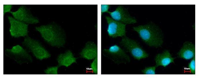
14-3-3 sigma antibody [C1C3] detects 14-3-3 sigma protein at cytoplasm by immunofluorescent analysis. Sample: A431 cells were fixed in ice-cold Methanol for 5 min. Green: 14-3-3 sigma protein stained by 14-3-3 sigma antibody [C1C3] diluted at 1:500. Blue: Hoechst 33343 staining.Application
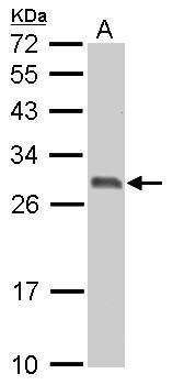
14-3-3 sigma antibody detects SFN protein by western blot analysis. A. 30 µg PC-12 whole cell lysate/extract 12% SDS-PAGE 14-3-3 sigma antibody dilution: 1:3000 The HRP-conjugated anti-rabbit IgG antibody was used to detect the primary antibody.Application
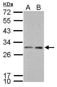
14-3-3 sigma antibody detects SFN protein by western blot analysis. A. 30 µg Neuro2A whole cell lysate/extract B. 30 µg GL261 whole cell lysate/extract 12% SDS-PAGE 14-3-3 sigma antibody dilution: 1:3000 The HRP-conjugated anti-rabbit IgG antibody was used to detect the primary antibody.Application
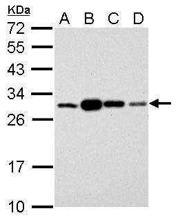
14-3-3 sigma antibody detects SFN protein by western blot analysis. A. 30 µg 293T whole cell lysate/extract B. 30 µg A431 whole cell lysate/extract C. 30 µg HeLa whole cell lysate/extract D. 30 µg HepG2 whole cell lysate/extract 12% SDS-PAGE 14-3-3 sigma antibody dilution: 1:3000 The HRP-conjugated anti-rabbit IgG antibody was used to detect the primary antibody.Application
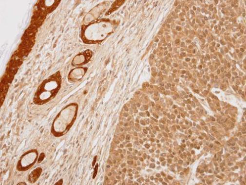
Immunohistochemical analysis of paraffin-embedded 59T xenograft, using 14-3-3 sigma antibody at 1:100 dilution.Application
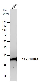
Whole cell extract (30 µg) was separated by 12% SDS-PAGE, and the membrane was blotted with 14-3-3 sigma antibody diluted at 1:3000. The HRP-conjugated anti-rabbit IgG antibody was used to detect the primary antibody.Application
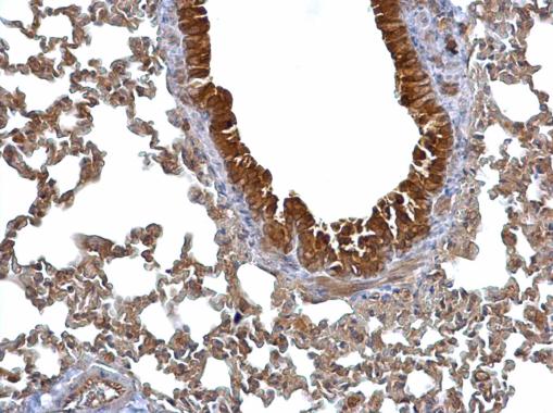
14-3-3 sigma antibody detects 14-3-3 sigma protein at cytosol on mouse lung by immunohistochemical analysis. Sample: Paraffin-embedded mouse lung. 14-3-3 sigma antibody dilution: 1:500.| Product Name | 14-3-3 sigma antibody |
|---|---|
| Antibody Type | Primary Antibodies |
| Product description | Rabbit Polyclonal antibody to 14-3-3 sigma (stratifin)Adapter protein implicated in the regulation of a large spectrum of both general and specialized signaling pathway. Binds to a large number of partners, usually by recognition of a phosphoserine or phosphothreonine motif. Binding generally results in the modulation of the activity of the binding partner. When bound to KRT17, regulates protein synthesis and epithelial cell growth by stimulating Akt/mTOR pathway. p53-regulated inhibitor of G2/M progression. |
| Immunogen | Carrier-protein conjugated synthetic peptide encompassing a sequence within the C-terminus region of human 14-3-3 sigma. The exact sequence is proprietary. |
| Clonality | Polyclonal |
|---|---|
| Isotype | IgG |
| Host Species | Rabbit |
| Tested Applications | ICC/IFIHC-PWB |
| *Optimal dilutions/concentrations should be determined by the researcher.
: |
|
| Species Reactivity | HumanMouseRat |
| Predicted Reactivity | Pig, Chicken, Sheep, Rhesus Monkey, BovinePig (100%), Chicken (100%), Sheep (100%), Rhesus Monkey (100%), Bovine (100%) |
| Concentration | 1mg/ml (Please refer to the vial label for the specific concentration) |
| Purification | Affinity purified |
| Gene Synonyms | YWHAS antibody SFN antibody epithelial cell marker protein 1 antibody 14-3-3 protein sigma antibody 14-3-3 sigma antibody stratifin antibody |
|---|---|
| Molecular Weight(MW) | 28 kDa (note) |
| Cellular Localization | Cytoplasm , Nucleus , Secreted |
Application
14-3-3 sigma antibody [C1C3] detects 14-3-3 sigma protein at cytoplasm by immunofluorescent analysis. Sample: A431 cells were fixed in ice-cold Methanol for 5 min. Green: 14-3-3 sigma protein stained by 14-3-3 sigma antibody [C1C3] diluted at 1:500. Blue: Hoechst 33343 staining.
Application
14-3-3 sigma antibody detects SFN protein by western blot analysis. A. 30 µg PC-12 whole cell lysate/extract 12% SDS-PAGE 14-3-3 sigma antibody dilution: 1:3000 The HRP-conjugated anti-rabbit IgG antibody was used to detect the primary antibody.
Application
14-3-3 sigma antibody detects SFN protein by western blot analysis. A. 30 µg Neuro2A whole cell lysate/extract B. 30 µg GL261 whole cell lysate/extract 12% SDS-PAGE 14-3-3 sigma antibody dilution: 1:3000 The HRP-conjugated anti-rabbit IgG antibody was used to detect the primary antibody.
Application
14-3-3 sigma antibody detects SFN protein by western blot analysis. A. 30 µg 293T whole cell lysate/extract B. 30 µg A431 whole cell lysate/extract C. 30 µg HeLa whole cell lysate/extract D. 30 µg HepG2 whole cell lysate/extract 12% SDS-PAGE 14-3-3 sigma antibody dilution: 1:3000 The HRP-conjugated anti-rabbit IgG antibody was used to detect the primary antibody.
Application
Immunohistochemical analysis of paraffin-embedded 59T xenograft, using 14-3-3 sigma antibody at 1:100 dilution.
Application
Whole cell extract (30 µg) was separated by 12% SDS-PAGE, and the membrane was blotted with 14-3-3 sigma antibody diluted at 1:3000. The HRP-conjugated anti-rabbit IgG antibody was used to detect the primary antibody.
Application
14-3-3 sigma antibody detects 14-3-3 sigma protein at cytosol on mouse lung by immunohistochemical analysis. Sample: Paraffin-embedded mouse lung. 14-3-3 sigma antibody dilution: 1:500.| Positive Control | 293T , A431 , HeLa , HepG2 , Neuro2A , GL261 , PC-12 , A549 |
|---|---|
| Application Notes | *Optimal dilutions/concentrations should be determined by the researcher.
: |
| Form | Liquid |
|---|---|
| Storage Instructions | Keep as concentrated solution. Aliquot and store at -20ºC or below. Avoid multiple freeze-thaw cycles. |
| Storage Buffer | 1XPBS, 1% BSA, 20% Glycerol (pH7). 0.01% Thimerosal was added as a preservative. |


