Application
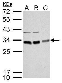
BCL10 antibody detects BCL10 protein by Western blot analysis. A. 30 µg Neuro2A whole cell lysate/extract B. 30 µg GL261 whole cell lysate/extract C. 30 µg C8D30 whole cell lysate/extract 12 % SDS-PAGE BCL10 antibody dilution: 1:1000Application
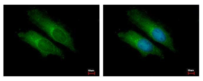
BCL10 antibody detects BCL10 protein at cytoplasm by immunofluorescent analysis. Sample: HeLa cells were fixed in 2% paraformaldehyde/culture medium at 37℃ for 30 min. Green: BCL10 protein stained by BCL10 antibody diluted at 1:500. Blue: Hoechst 33342 staining.Application
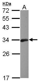
Sample (30 ug of whole cell lysate) A: Raji 12% SDS PAGE diluted at 1:1000Application
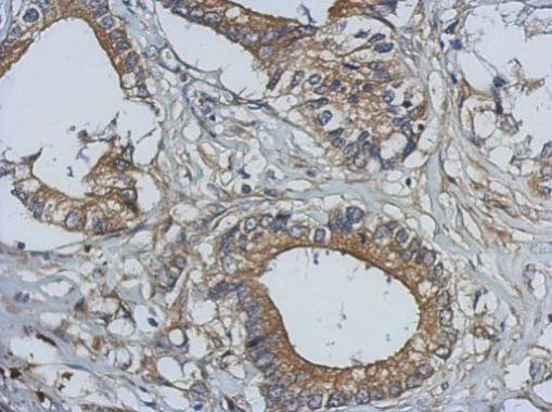
Immunohistochemical analysis of paraffin-embedded human gastric cancer, using BCL10 antibody antibody at 1:500 dilution.Application
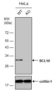
Wild-type (WT) and BCL10 knockout (KO) HeLa cell extracts (30 µg) were separated by 12% SDS-PAGE, and the membrane was blotted with BCL10 antibody diluted at 1:2500. The HRP-conjugated anti-rabbit IgG antibody was used to detect the primary antibody.Application
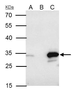
BCL10 antibody immunoprecipitates BCL10 protein in IP experiments. IP samples: Raji whole cell extract A. 40 μg Raji whole cell extract B. Control with 4 μg of preimmune Rabbit IgG C. Immunoprecipitation of BCL10 protein by 4 μg BCL10 antibody 12 % SDS-PAGE The immunoprecipitated BCL10 protein was detected by BCL10 antibody diluted at 1:500. [EasyBlot anti-rabbit IgG was used as a secondary reagent]| Product Name | BCL10 antibody |
|---|---|
| Antibody Type | Primary Antibodies |
| Product description | Rabbit Polyclonal antibody to BCL10 (B-cell CLL/lymphoma 10)This gene was identified by its translocation in a case of mucosa-associated lymphoid tissue (MALT) lymphoma. The protein encoded by this gene contains a caspase recruitment domain (CARD), and has been shown to induce apoptosis and to activate NF-kappaB. This protein is reported to interact with other CARD domain containing proteins including CARD9, 10, 11 and 14, which are thought to function as upstream regulators in NF-kappaB signaling. This protein is found to form a complex with MALT1, a protein encoded by another gene known to be translocated in MALT lymphoma. MALT1 and this protein are thought to synergize in the activation of NF-kappaB, and the deregulation of either of them may contribute to the same pathogenetic process that leads to the malignancy. [provided by RefSeq] |
| Immunogen | Recombinant protein encompassing a sequence within the center region of human BCL10. The exact sequence is proprietary. |
| Clonality | Polyclonal |
|---|---|
| Isotype | IgG |
| Host Species | Rabbit |
| Tested Applications | ICC/IFIHC-PIPWB |
| *Optimal dilutions/concentrations should be determined by the researcher.
: |
|
| Species Reactivity | HumanMouse |
| Predicted Reactivity | Rat, Pig, Rhesus Monkey, BovineRat (92%), Pig (92%), Rhesus Monkey (99%), Bovine (95%) |
| Concentration | 1mg/ml (Please refer to the vial label for the specific concentration) |
| Purification | Affinity purified |
| Gene Synonyms | CARMEN antibody CIPER antibody CLAP antibody c-E10 antibody mE10 antibody BCL10 antibody B-cell lymphoma/leukemia 10 antibody cellular-E10 antibody cCARMEN antibody CARD containing molecule enhancing NF-kB antibody hCLAP antibody CED-3/ICH-1 pro |
|---|---|
| Molecular Weight(MW) | 26 kDa (note) (2) |
| Cellular Localization | Cytoplasm , perinuclear region |
Application
BCL10 antibody detects BCL10 protein by Western blot analysis. A. 30 µg Neuro2A whole cell lysate/extract B. 30 µg GL261 whole cell lysate/extract C. 30 µg C8D30 whole cell lysate/extract 12 % SDS-PAGE BCL10 antibody dilution: 1:1000
Application
BCL10 antibody detects BCL10 protein at cytoplasm by immunofluorescent analysis. Sample: HeLa cells were fixed in 2% paraformaldehyde/culture medium at 37℃ for 30 min. Green: BCL10 protein stained by BCL10 antibody diluted at 1:500. Blue: Hoechst 33342 staining.
Application
Sample (30 ug of whole cell lysate) A: Raji 12% SDS PAGE diluted at 1:1000
Application
Immunohistochemical analysis of paraffin-embedded human gastric cancer, using BCL10 antibody antibody at 1:500 dilution.
Application
Wild-type (WT) and BCL10 knockout (KO) HeLa cell extracts (30 µg) were separated by 12% SDS-PAGE, and the membrane was blotted with BCL10 antibody diluted at 1:2500. The HRP-conjugated anti-rabbit IgG antibody was used to detect the primary antibody.
Application
BCL10 antibody immunoprecipitates BCL10 protein in IP experiments. IP samples: Raji whole cell extract A. 40 μg Raji whole cell extract B. Control with 4 μg of preimmune Rabbit IgG C. Immunoprecipitation of BCL10 protein by 4 μg BCL10 antibody 12 % SDS-PAGE The immunoprecipitated BCL10 protein was detected by BCL10 antibody diluted at 1:500. [EasyBlot anti-rabbit IgG was used as a secondary reagent]| Positive Control | Raji , Neuro2A , GL261 , C8D30 |
|---|---|
| Application Notes | *Optimal dilutions/concentrations should be determined by the researcher.
: |
| Form | Liquid |
|---|---|
| Storage Instructions | Keep as concentrated solution. Aliquot and store at -20ºC or below. Avoid multiple freeze-thaw cycles. |
| Storage Buffer | 1XPBS, 1% BSA, 20% Glycerol (pH7). 0.01% Thimerosal was added as a preservative. |


