Application
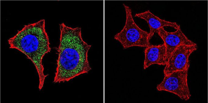
Immunofluorescent analysis of beta Catenin in HeLa cells. beta Catenin staining (green), F-Actin staining with Phalloidin (red) and nuclei with DAPI (blue) is shown. Cells were grown on slides and fixed with formaldehyde prior to staining. Cells were probed without (control) or with beta Catenin antibody [9F2] at a dilution of 1:20 over night at 4 °C, washed with PBS and incubated with a proper secondary antibody. Images were taken at 60X magnification.| Product Name | beta Catenin antibody [9F2] |
|---|---|
| Antibody Type | Primary Antibodies |
| Product description | Mouse monoclonal antibody [9F2] to beta CateninThe protein encoded by this gene is part of a complex of proteins that constitute adherens junctions (AJs). AJs are necessary for the creation and maintenance of epithelial cell layers by regulating cell growth and adhesion between cells. The encoded protein also anchors the actin cytoskeleton and may be responsible for transmitting the contact inhibition signal that causes cells to stop dividing once the epithelial sheet is complete. Finally, this protein binds to the product of the APC gene, which is mutated in adenomatous polyposis of the colon. Mutations in this gene are a cause of colorectal cancer (CRC), pilomatrixoma (PTR), medulloblastoma (MDB), and ovarian cancer. Three transcript variants encoding the same protein have been found for this gene.[provided by RefSeq, Oct 2009] |
| Immunogen | 120 kDa MBP/beta catenin fusion protein purified on amylose column. |
| Clonality | Monoclonal |
|---|---|
| Isotype | IgG1 |
| Clone Number | 9F2 |
| Host Species | Mouse |
| Tested Applications | ICC/IFIPWB |
| ICC/IF: 10 ug/ml | |
| Species Reactivity | HumanMouse |
| Concentration | 1mg/ml (Please refer to the vial label for the specific concentration) |
| Purification | Protein G |
| Gene Synonyms | armadillo , MRD19 , CTNNB , CTNNB1 , catenin (cadherin-associated protein), beta 1, 88kDa |
|---|---|
| Target | beta Catenin |
Application
Immunofluorescent analysis of beta Catenin in HeLa cells. beta Catenin staining (green), F-Actin staining with Phalloidin (red) and nuclei with DAPI (blue) is shown. Cells were grown on slides and fixed with formaldehyde prior to staining. Cells were probed without (control) or with beta Catenin antibody [9F2] at a dilution of 1:20 over night at 4 °C, washed with PBS and incubated with a proper secondary antibody. Images were taken at 60X magnification.| Application Notes | ICC/IF: 10 ug/ml |
|---|
| Form | Liquid |
|---|---|
| Storage Instructions | -20° C, Avoid Freeze/Thaw Cycles |
| Storage Buffer | PBS containing 0.05 % sodium azide and 1 mg/ml BSA |


