Application
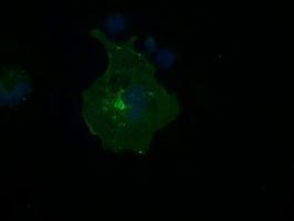
Anti-FCGR2A mouse monoclonal antibody immunofluorescent staining of COS7 cells transiently transfected with FCGR2AApplication
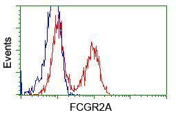
HEK293T cells transfected with either pCMV6-ENTRY FCGR2AApplication
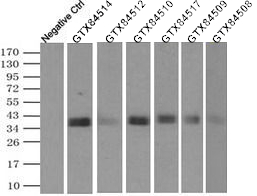
IP was performed using 500ul of DDDDK tagged FCGR2A overexpression lysates and 2ug of CD32a antibody. The precipitates were detected by DDDDK tag antibody.Application
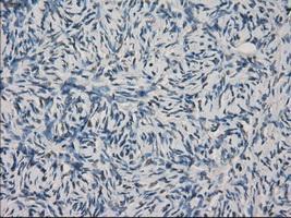
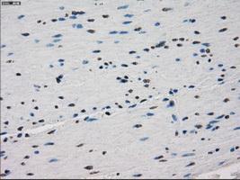
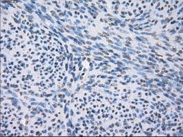
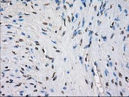
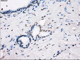
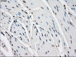
Immunohistochemical staining of paraffin-embedded Ovary tissue using anti-FCGR2A mouse monoclonal antibody. ( Dilution 1:50)Application
Immunohistochemical staining of paraffin-embedded colon tissue using anti-FCGR2A mouse monoclonal antibody. ( Dilution 1:50)Application
Immunohistochemical staining of paraffin-embedded endometrium tissue using anti-FCGR2A mouse monoclonal antibody. ( Dilution 1:50)Application
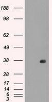
HEK293T cells were transfected with the pCMV6-ENTRY control (Left lane) or pCMV6-ENTRY FCGR2A (Right lane) cDNA for 48 hrs and lysed. Equivalent amounts of cell lysates (5 ug per lane) were separated by SDS-PAGE and immunoblotted with anti-FCGR2A .Application
Immunohistochemical staining of paraffin-embedded prostate tissue using anti-FCGR2A mouse monoclonal antibody. ( Dilution 1:50)Application
Immunohistochemical staining of paraffin-embedded pancreas tissue using anti-FCGR2A mouse monoclonal antibody. ( Dilution 1:50)Application
Immunohistochemical staining of paraffin-embedded bladder tissue using anti-FCGR2A mouse monoclonal antibody. ( Dilution 1:50)| Product Name | CD32a antibody |
|---|---|
| Antibody Type | Primary Antibodies |
| Product description | Mouse monoclonal antibody [16D6] to CD32aBinds to the Fc region of immunoglobulins gamma. Low affinity receptor. By binding to IgG it initiates cellular responses against pathogens and soluble antigens. |
| Immunogen | Protein expressed in 293T cell transfected with human FCGR2A expression vector |
| Clonality | Monoclonal |
|---|---|
| Isotype | IgG1 |
| Clone Number | 16D6 |
| Host Species | Mouse |
| Tested Applications | FACSICC/IFIHC-PIPWB |
| We recommend the following starting dilutions:
For WB: Use at a dilution of 1:200-1:1000.
For IHC-P: Use at a dilution of 1:50.
For ICC/IF: Use at a dilution of 1:100.
For FACS: Use at a dilution of 1:100.
For IP: Use at a concentration of 2ug/500ul.
Optimal dilutions should be determined experimentally by the end user.: |
|
| Species Reactivity | Human |
| Concentration | 1mg/ml |
| Purification | Affinity purified |
| Gene Synonyms | FcGR IGFR2 CD32 FCG2 FCGR2 MGC30032 CDw32 FCGR2A1 FCGR2A MGC23887 |
|---|---|
| Target | CD32a |
Application
Anti-FCGR2A mouse monoclonal antibody immunofluorescent staining of COS7 cells transiently transfected with FCGR2A
Application
HEK293T cells transfected with either pCMV6-ENTRY FCGR2A
Application
IP was performed using 500ul of DDDDK tagged FCGR2A overexpression lysates and 2ug of CD32a antibody. The precipitates were detected by DDDDK tag antibody.
Application
Immunohistochemical staining of paraffin-embedded Ovary tissue using anti-FCGR2A mouse monoclonal antibody. ( Dilution 1:50)
Application
Immunohistochemical staining of paraffin-embedded colon tissue using anti-FCGR2A mouse monoclonal antibody. ( Dilution 1:50)
Application
Immunohistochemical staining of paraffin-embedded endometrium tissue using anti-FCGR2A mouse monoclonal antibody. ( Dilution 1:50)
Application
HEK293T cells were transfected with the pCMV6-ENTRY control (Left lane) or pCMV6-ENTRY FCGR2A (Right lane) cDNA for 48 hrs and lysed. Equivalent amounts of cell lysates (5 ug per lane) were separated by SDS-PAGE and immunoblotted with anti-FCGR2A .
Application
Immunohistochemical staining of paraffin-embedded prostate tissue using anti-FCGR2A mouse monoclonal antibody. ( Dilution 1:50)
Application
Immunohistochemical staining of paraffin-embedded pancreas tissue using anti-FCGR2A mouse monoclonal antibody. ( Dilution 1:50)
Application
Immunohistochemical staining of paraffin-embedded bladder tissue using anti-FCGR2A mouse monoclonal antibody. ( Dilution 1:50)| Application Notes | We recommend the following starting dilutions:
For WB: Use at a dilution of 1:200-1:1000.
For IHC-P: Use at a dilution of 1:50.
For ICC/IF: Use at a dilution of 1:100.
For FACS: Use at a dilution of 1:100.
For IP: Use at a concentration of 2ug/500ul.
Optimal dilutions should be determined experimentally by the end user.: |
|---|
| Form | Liquid |
|---|---|
| Storage Instructions | For short-term storage, store at 4°C or aliquot into working amounts and store at -20°C. For long-term storage, store at -70°C (aliquotted). Avoid repeated freeze-thaw cycles. |
| Storage Buffer | PBS (PH 7.3) containing 1% BSA, 50% glycerol and 0.02% sodium azide. |


