Application
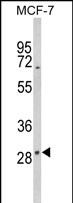
Western blot analysis of DIO2 Antibody (Center) in MCF-7 cell line lysates (35ug/lane). DIO2 (arrow) was detected using the purified Pab.Application
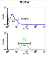
DIO2 Antibody (Center) flow cytometric analysis of MCF-7 cells (bottom histogram) compared to a negative control cell (top histogram).FITC-conjugated goat-anti-rabbit secondary antibodies were used for the analysis.Application
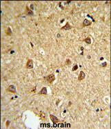
DIO2 Antibody (Center) IHC analysis in formalin fixed and paraffin embedded mouse brain followed by peroxidase conjugation of the secondary antibody and DAB staining. This data demonstrates the use of the mouse DIO2 Antibody (Center) for immunohistochemistry. Clinical relevance has not been evaluated.| Product Name | DIO2 antibody, Internal |
|---|---|
| Antibody Type | Primary Antibodies |
| Product description | Rabbit polyclonal to DIO2 (Internal)The protein encoded by this gene belongs to the iodothyronine deiodinase family. It activates thyroid hormone by converting the prohormone thyroxine (T4) by outer ring deiodination (ORD) to bioactive 3,3',5-triiodothyronine (T3). It is highly expressed in the thyroid, and may contribute significantly to the relative increase in thyroidal T3 production in patients with Graves disease and thyroid adenomas. This protein contains selenocysteine (Sec) residues encoded by the UGA codon, which normally signals translation termination. The 3' UTR of Sec-containing genes have a common stem-loop structure, the sec insertion sequence (SECIS), which is necessary for the recognition of UGA as a Sec codon rather than as a stop signal. Alternative splicing results in multiple transcript variants encoding different isoforms. [provided by RefSeq] |
| Immunogen | KLH conjugated synthetic peptide between 171~200 amino acids from the Center region of human DIO2. |
| Clonality | Polyclonal |
|---|---|
| Isotype | IgG |
| Host Species | Rabbit |
| Tested Applications | FACSIHC-PWB |
| ELISA: Use at 1:1,000: |
|
| Species Reactivity | Human |
| Concentration | 0.2mg/ml |
| Purification | Affinity purified |
| Gene Synonyms | DIO2 Type2 DI TypeII5'deiodinase 5DII TypeII 5'deiodinase Type 2 DI DIOII ITDI2 Type-II 5'-deiodinase Type II iodothyronine deiodinase TXDI2 Type II 5' deiodinase |
|---|---|
| Target | DIO2 |
| Molecular Weight(MW) | 30.552 kDa (note) |
Application
Western blot analysis of DIO2 Antibody (Center) in MCF-7 cell line lysates (35ug/lane). DIO2 (arrow) was detected using the purified Pab.
Application
DIO2 Antibody (Center) flow cytometric analysis of MCF-7 cells (bottom histogram) compared to a negative control cell (top histogram).FITC-conjugated goat-anti-rabbit secondary antibodies were used for the analysis.
Application
DIO2 Antibody (Center) IHC analysis in formalin fixed and paraffin embedded mouse brain followed by peroxidase conjugation of the secondary antibody and DAB staining. This data demonstrates the use of the mouse DIO2 Antibody (Center) for immunohistochemistry. Clinical relevance has not been evaluated.| Application Notes | ELISA: Use at 1:1,000: |
|---|
| Form | Liquid |
|---|---|
| Storage Instructions | Store at at 2-8°C for up to 6 months. For long term storage, aliquot and store at -20 °C. Avoid repeated freeze/thaw cycles. |
| Storage Buffer | PBS with 0.09% (W/V) sodium azide |


