Application
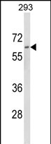
Western blot analysis of DOLK Antibody (Center) in 293 cell line lysates (35ug/lane). DOLK (arrow) was detected using the purified Pab.Application
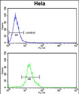
DOLK Antibody (Center) flow cytometry analysis of Hela cells (bottom histogram) compared to a negative control cell (top histogram).FITC-conjugated goat-anti-rabbit secondary antibodies were used for the analysis.Application
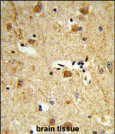
Formalin-fixed and paraffin-embedded human brain tissue reacted with DOLK Antibody (Center), which was peroxidase-conjugated to the secondary antibody, followed by DAB staining. This data demonstrates the use of this antibody for immunohistochemistry; clinical relevance has not been evaluated.| Product Name | DOLK antibody, Internal |
|---|---|
| Antibody Type | Primary Antibodies |
| Product description | Rabbit polyclonal to DOLK (Internal)The protein encoded by this gene catalyzes the CTP-mediated phosphorylation of dolichol, and is involved in the synthesis of Dol-P-Man, which is an essential glycosyl carrier lipid for C- and O-mannosylation, N- and O-linked glycosylation of proteins, and for the biosynthesis of glycosyl phosphatidylinositol anchors in endoplasmic reticulum. Mutations in this gene are associated with dolichol kinase deficiency. |
| Immunogen | KLH conjugated synthetic peptide between 318~348 amino acids from the Center region of human DOLK. |
| Clonality | Polyclonal |
|---|---|
| Isotype | IgG |
| Host Species | Rabbit |
| Tested Applications | FACSIHC-PWB |
| ELISA: Use at 1:1,000: |
|
| Species Reactivity | Human |
| Concentration | 0.2mg/ml |
| Purification | Affinity purified |
| Gene Synonyms | DOLK DOLK KIAA1094 TMEM15 UNQ2422/PRO4980 Transmembrane protein15 Transmembrane protein 15 Dolichol kinase |
|---|---|
| Molecular Weight(MW) | 59.268 kDa (note) |
Application
Western blot analysis of DOLK Antibody (Center) in 293 cell line lysates (35ug/lane). DOLK (arrow) was detected using the purified Pab.
Application
DOLK Antibody (Center) flow cytometry analysis of Hela cells (bottom histogram) compared to a negative control cell (top histogram).FITC-conjugated goat-anti-rabbit secondary antibodies were used for the analysis.
Application
Formalin-fixed and paraffin-embedded human brain tissue reacted with DOLK Antibody (Center), which was peroxidase-conjugated to the secondary antibody, followed by DAB staining. This data demonstrates the use of this antibody for immunohistochemistry; clinical relevance has not been evaluated.| Application Notes | ELISA: Use at 1:1,000: |
|---|
| Form | Liquid |
|---|---|
| Storage Instructions | Store at at 2-8°C for up to 6 months. For long term storage, aliquot and store at -20 °C. Avoid repeated freeze/thaw cycles. |
| Storage Buffer | PBS with 0.09% (W/V) sodium azide |


