Application
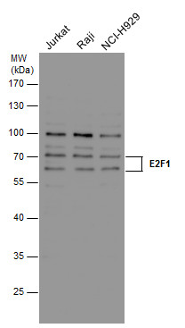
E2F1 antibody [16G7] detects E2F1 protein by western blot analysis. Various whole cell extracts (30 μg) were separated by 10% SDS-PAGE, and the membrane was blotted with E2F1 antibody [16G7] diluted at a dilution of 1:1000.Application
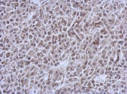
Immunohistochemical analysis of paraffin-embedded HBL435 xenograft, using E2F1 antibody at 1:200 dilution.Application
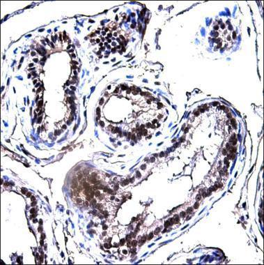
Immunohistochemical analysis of paraffin-embedded Mouse prostate, using E2F1 antibody at 1:150 dilution. Image courtesy of Ali Kuraishy Postdoc (Laboratory of Dr. Michael Karin, UCSD.)Application
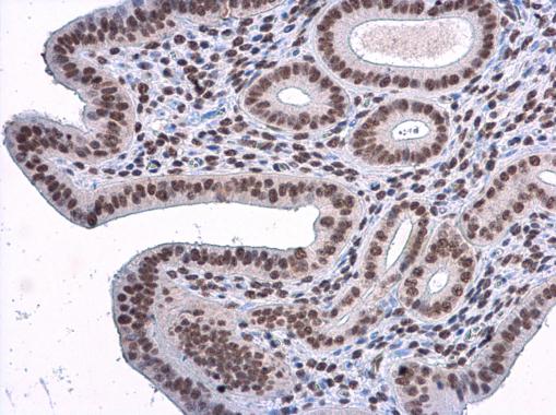
E2F1 antibody [16G7] detects E2F1 protein at nucleus in mouse cervix by immunohistochemical analysis. Sample: Paraffin-embedded mouse cervix. E2F1 antibody [16G7] diluted at 1:500.Application
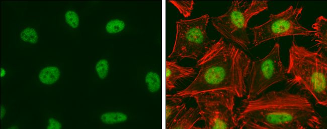
E2F1 antibody [16G7] detects E2F1 protein at nucleus by immunofluorescent analysis. Sample: HeLa cells were fixed in 4% paraformaldehyde at RT for 15 min. Green: E2F1 protein stained by E2F1 antibody [16G7] diluted at 1:200. Red: phalloidin, a cytoskeleton marker, diluted at 1:50.WB
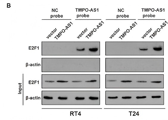
(B) An RNA pull-down assay was performed to validate the potential interaction between TMPO-AS1 and E2F1 in RT4 and T24 cells overexpressing TMPO-AS1.WB
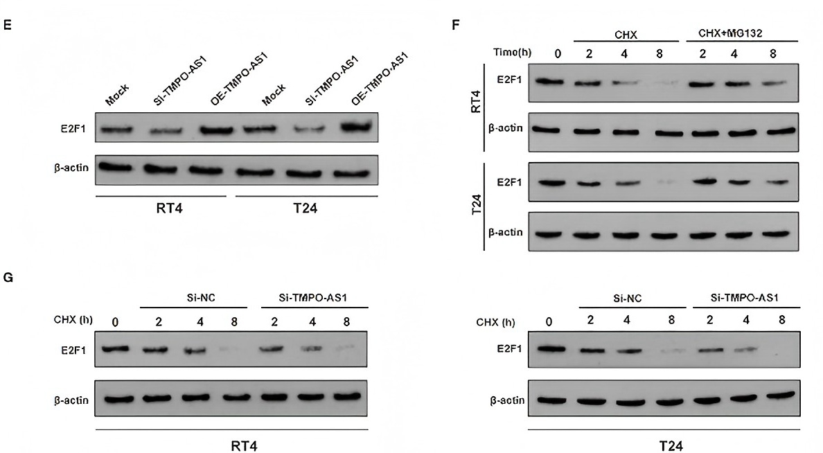
(E) The E2F1 protein expression was evaluated via Western blotting in TMPO-AS1 knockout or overexpressing RT4 and T24 cells. (F) Western blotting show that the proteasome inhibitor MG132 protein increased the stability of E2F1 in cycloheximide-treated RT4 and T24 cells. (G) Western blotting showing the decreased E2F1 stability in TMPO-AS1 knockdown RT4 and T24 cells.WB
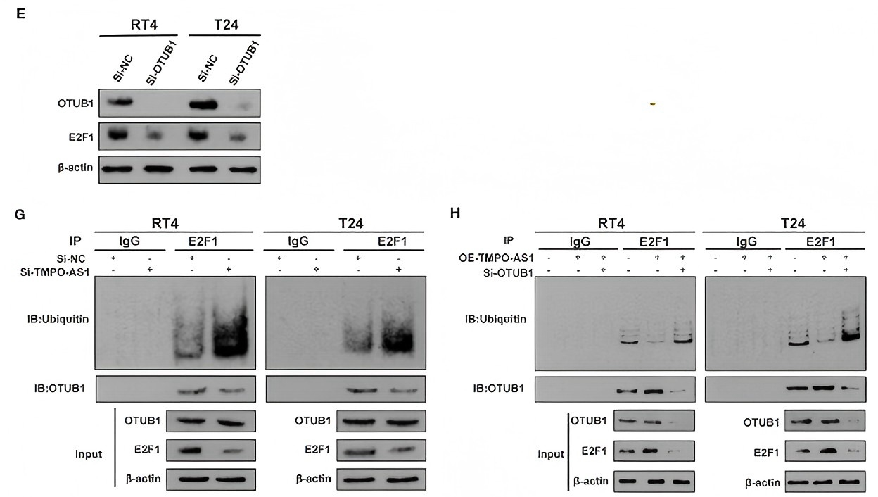
(E) Western blotting images showing that the knockdown of OTUB1 leads to the decrease in the E2F1 protein levels in RT4 and T24 cells. Si- OTUB1, siRNA targeting OTUB1.(G) Co-IP assays were performed in control or si-TMPO-AS1-treated RT4 and T24 cells using an anti-E2F1 antibody, followed by Western blotting to analyze the ubiquitin levels of E2F1. (H) RT4 and T24 cells transfected with the empty vector, and the construct for the overexpression of TMPO-AS1, alone, or together with si-OTUB1 were subjected to immunoprecipitation with an anti-E2F1 antibody, followed by Western blotting to analyze the ubiquitin levels of E2F1.WB
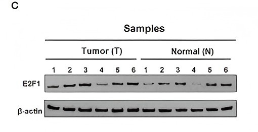
(C) The protein expression of E2F1 was assessed via Western blotting in six paired BC and adjacent normal tissue samples.IHC
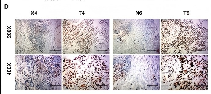
(D) The protein expression of E2F1 was also assessed using immunohistochemistry in six paired BC and adjacent normal tissue samples.| Product Name | E2F1 antibody |
|---|---|
| Antibody Type | Primary Antibodies |
| Product description | Mouse monoclonal [16G7] antibody to E2F - 1E2F-1-16G7 recognizes E2F-1 that was identified as a cellular protein with DNA binding activity associated with the adenovirus E2 gene promoter. The original member of the E2F family of transcription factors, now consisting of five, was identified through its association with the retinoblastoma protein, which controls its activity. E2F-1 is cell cycle regulated with very low levels in early G1 then increasing levels as cells move from G1 to S with highest levels of protein at the G1/S boundary, which is consistent with its role in S-phase entry. |
| Immunogen | The region encoding the DNA binding domain (amino acids 244-264) of E2F-1 expressed in E. coli. |
| Clonality | Monoclonal |
|---|---|
| Isotype | IgG1 |
| Clone Number | 16G7 |
| Host Species | Mouse |
| Tested Applications | ICC/IFIHC-PIPWB |
| *Optimal dilutions/concentrations should be determined by the researcher.
: |
|
| Species Reactivity | HumanMouse |
| Concentration | 1mg/ |
| Purification | Affinity purified |
| Gene Synonyms | E2F1 E2F-1 Q01094 189971 1869 RBP3 RBBP3 RBAP1 E2F 1 |
|---|---|
| Target | E2F1 |
| Cellular Localization | Nuclear |
| Light Chain Type | kappa |
Application
E2F1 antibody [16G7] detects E2F1 protein by western blot analysis. Various whole cell extracts (30 μg) were separated by 10% SDS-PAGE, and the membrane was blotted with E2F1 antibody [16G7] diluted at a dilution of 1:1000.
Application
Immunohistochemical analysis of paraffin-embedded HBL435 xenograft, using E2F1 antibody at 1:200 dilution.
Application
Immunohistochemical analysis of paraffin-embedded Mouse prostate, using E2F1 antibody at 1:150 dilution. Image courtesy of Ali Kuraishy Postdoc (Laboratory of Dr. Michael Karin, UCSD.)
Application
E2F1 antibody [16G7] detects E2F1 protein at nucleus in mouse cervix by immunohistochemical analysis. Sample: Paraffin-embedded mouse cervix. E2F1 antibody [16G7] diluted at 1:500.
Application
E2F1 antibody [16G7] detects E2F1 protein at nucleus by immunofluorescent analysis. Sample: HeLa cells were fixed in 4% paraformaldehyde at RT for 15 min. Green: E2F1 protein stained by E2F1 antibody [16G7] diluted at 1:200. Red: phalloidin, a cytoskeleton marker, diluted at 1:50.
WB
(B) An RNA pull-down assay was performed to validate the potential interaction between TMPO-AS1 and E2F1 in RT4 and T24 cells overexpressing TMPO-AS1.
WB
(E) The E2F1 protein expression was evaluated via Western blotting in TMPO-AS1 knockout or overexpressing RT4 and T24 cells. (F) Western blotting show that the proteasome inhibitor MG132 protein increased the stability of E2F1 in cycloheximide-treated RT4 and T24 cells. (G) Western blotting showing the decreased E2F1 stability in TMPO-AS1 knockdown RT4 and T24 cells.
WB
(E) Western blotting images showing that the knockdown of OTUB1 leads to the decrease in the E2F1 protein levels in RT4 and T24 cells. Si- OTUB1, siRNA targeting OTUB1.(G) Co-IP assays were performed in control or si-TMPO-AS1-treated RT4 and T24 cells using an anti-E2F1 antibody, followed by Western blotting to analyze the ubiquitin levels of E2F1. (H) RT4 and T24 cells transfected with the empty vector, and the construct for the overexpression of TMPO-AS1, alone, or together with si-OTUB1 were subjected to immunoprecipitation with an anti-E2F1 antibody, followed by Western blotting to analyze the ubiquitin levels of E2F1.
WB
(C) The protein expression of E2F1 was assessed via Western blotting in six paired BC and adjacent normal tissue samples.
IHC
(D) The protein expression of E2F1 was also assessed using immunohistochemistry in six paired BC and adjacent normal tissue samples.| Positive Control | Jurkat , Raji , NCI-H929 |
|---|---|
| Application Notes | *Optimal dilutions/concentrations should be determined by the researcher.
: |
| Form | Liquid |
|---|---|
| Storage Instructions | Keep as concentrated solution. Store at 4°C short term. For extended storage aliquot and store at -20°C or below. Avoid freeze-thaw cycles. |
| Storage Buffer | Phosphate-buffered saline, pH 7.4, containing no preservatives |


