Application
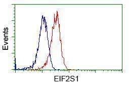
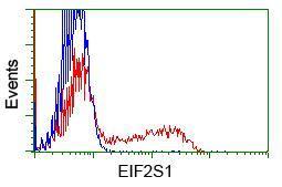
Flow cytometric Analysis of Jurkat cells, using anti-EIF2S1 antibody,(Red), compared to a nonspecific negative control antibody(TA50011),(Blue).Application
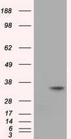
HEK293T cells were transfected with the pCMV6-ENTRY control (Left lane) or pCMV6-ENTRY EIF2S1 (Right lane) cDNA for 48 hrs and lysed. Equivalent amounts of cell lysates (5 ug per lane) were separated by SDS-PAGE and immunoblotted with anti-EIF2S1.Application
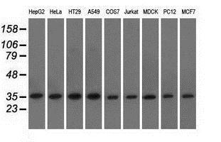
Western blot analysis of extracts (35ug) from 9 different cell lines by using anti-EIF2S1 monoclonal antibody.Application
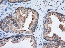
Immunohistochemical staining of paraffin-embedded Human prostate tissue using anti-EIF2S1 mouse monoclonal antibody. ( Dilution 1:50)Application
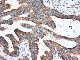
Immunohistochemical staining of paraffin-embedded Adenocarcinoma of Human colon tissue using anti-EIF2S1 mouse monoclonal antibody. ( Dilution 1:50)Application
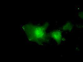
Anti-EIF2S1 mouse monoclonal antibody immunofluorescent staining of COS7 cells transiently transfected with EIF2S1Application
HEK293T cells transfected with either RC200368 overexpress plasmid(Red) or empty vector control plasmid(Blue) were immunostained by anti-EIF2S1 antibody, and then analyzed by flow cytometry.Application
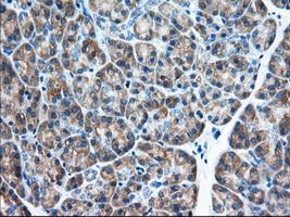
Immunohistochemical staining of paraffin-embedded Human pancreas tissue using anti-EIF2S1 mouse monoclonal antibody. ( Dilution 1:50)Application
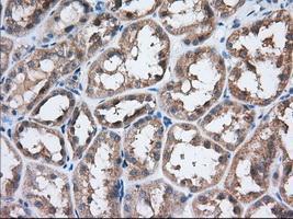
Immunohistochemical staining of paraffin-embedded Human Kidney tissue using anti-EIF2S1 mouse monoclonal antibody. ( Dilution 1:50)Application
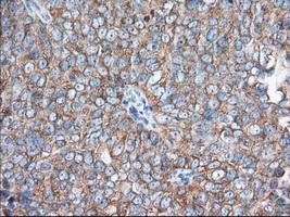
Immunohistochemical staining of paraffin-embedded Adenocarcinoma of Human ovary tissue using anti-EIF2S1 mouse monoclonal antibody. ( Dilution 1:50)| Product Name | eIF2 alpha antibody |
|---|---|
| Antibody Type | Primary Antibodies |
| Product description | Mouse monoclonal antibody [3H4] to eIF2 alphaFunctions in the early steps of protein synthesis by forming a ternary complex with GTP and initiator tRNA. This complex binds to a 40S ribosomal subunit, followed by mRNA binding to form a 43S preinitiation complex. Junction of the 60S ribosomal subunit to form the 80S initiation complex is preceded by hydrolysis of the GTP bound to eIF-2 and release of an eIF-2-GDP binary complex. In order for eIF-2 to recycle and catalyze another round of initiation, the GDP bound to eIF-2 must exchange with GTP by way of a reaction catalyzed by eIF-2B. |
| Immunogen | Full length human recombinant protein of human EIF2S1 (NP_004085) produced in HEK293T cell. |
| Clonality | Monoclonal |
|---|---|
| Isotype | IgG1 |
| Clone Number | 3H4 |
| Host Species | Mouse |
| Tested Applications | FACSICC/IFIHC-PWB |
| Recommended Starting Dilutions:
For WB: Use at a dilution of 1:1000-1:2000.
For IHC-P: Use at a dilution of 1:50.
For ICC/IF: Use at a dilution of 1:100.
For FACS: Use at a dilution of 1:100.
Optimal dilutions should be determined experimentally by the researcher.: |
|
| Species Reactivity | HumanDogMonkeyRat |
| Concentration | 1mg/ml |
| Purification | Affinity purified |
| Gene Synonyms | EIF2S1 EIF2 EIF-2 EIF-2alpha EIF2A EIF-2A EIF2alpha EIF 2 EIF 2A EIF 2alpha |
|---|---|
| Target | eIF2 alpha |
| Tissue Specificity | is specific for human EIF2S1 (NP_004085). |
Application
Flow cytometric Analysis of Jurkat cells, using anti-EIF2S1 antibody,(Red), compared to a nonspecific negative control antibody(TA50011),(Blue).
Application
HEK293T cells were transfected with the pCMV6-ENTRY control (Left lane) or pCMV6-ENTRY EIF2S1 (Right lane) cDNA for 48 hrs and lysed. Equivalent amounts of cell lysates (5 ug per lane) were separated by SDS-PAGE and immunoblotted with anti-EIF2S1.
Application
Western blot analysis of extracts (35ug) from 9 different cell lines by using anti-EIF2S1 monoclonal antibody.
Application
Immunohistochemical staining of paraffin-embedded Human prostate tissue using anti-EIF2S1 mouse monoclonal antibody. ( Dilution 1:50)
Application
Immunohistochemical staining of paraffin-embedded Adenocarcinoma of Human colon tissue using anti-EIF2S1 mouse monoclonal antibody. ( Dilution 1:50)
Application
Anti-EIF2S1 mouse monoclonal antibody immunofluorescent staining of COS7 cells transiently transfected with EIF2S1
Application
HEK293T cells transfected with either RC200368 overexpress plasmid(Red) or empty vector control plasmid(Blue) were immunostained by anti-EIF2S1 antibody, and then analyzed by flow cytometry.
Application
Immunohistochemical staining of paraffin-embedded Human pancreas tissue using anti-EIF2S1 mouse monoclonal antibody. ( Dilution 1:50)
Application
Immunohistochemical staining of paraffin-embedded Human Kidney tissue using anti-EIF2S1 mouse monoclonal antibody. ( Dilution 1:50)
Application
Immunohistochemical staining of paraffin-embedded Adenocarcinoma of Human ovary tissue using anti-EIF2S1 mouse monoclonal antibody. ( Dilution 1:50)| Application Notes | Recommended Starting Dilutions:
For WB: Use at a dilution of 1:1000-1:2000.
For IHC-P: Use at a dilution of 1:50.
For ICC/IF: Use at a dilution of 1:100.
For FACS: Use at a dilution of 1:100.
Optimal dilutions should be determined experimentally by the researcher.: |
|---|
| Form | Liquid |
|---|---|
| Storage Instructions | For short-term storage, store at 4°C or aliquot into working amounts and store at -20°C. For long-term storage, store at -70°C (aliquotted). Avoid repeated freeze-thaw cycles. |
| Storage Buffer | PBS (PH 7.3) containing 1% BSA, 50% glycerol and 0.02% sodium azide. |


