
Application
Blot of yeast protein extract stained with the anti-Gibrillarin anitbody, stained a single band at ~34kDa.Application
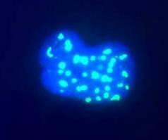
High magnification view of human Hek293 cell nuclei stained with mouse monoclonal to fibrillarin (green), counterstained with a fluorescent DNA probe (blue). Nuclear DNA is revealed with Hoechst dye (blue). Cultures were processed using our standard fixatApplication
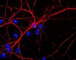
Rat neurons and glial stained with mouse monoclonal to Fibrillarin (green) and with chicken antibody to neurofilament NF-H (red). Cells were counterstained with a fluorescent DNA probe (blue). Nuclear DNA is revealed with Hoechst dye (blue).Application
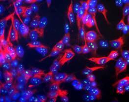
Human neuroblastoma line SH-SY5Y stained with mouse monoclonal to Fibrillarin (green) and with chicken antibody to neurofilament NF-H (red) and counterstained with a fluorescent DNA probe (blue). Nuclear DNA is revealed with Hoechst dye (blue). The NF-H antibody was used at a dilution of 1/100000 and the fibrillarin monoclonal at 1/1000| Product Name | Fibrillarin antibody |
|---|---|
| Antibody Type | Primary Antibodies |
| Product description | Mouse monoclonal [38F3] to Fibrillarin - Nucleolar MarkerNop1p / Fibrillarin was originally identified as a nucleolar protein of bakers yeast, Saccharomyces cerevisiae (accession P15646). The Nop1p protein is essential for yeast viability and is localized in the nucleoli. The human homologue of Nop1p is fibrillarin (accession P22087) a component of the nucleolar small nuclear ribonucleoprotein (snRNP) particle. The human fibrillarin gene is located on chromosome 19 (19q13.1). Fibrillarin proteins have been cloned and sequenced from several other species (Mouse, accession P35550, Xenopus accession P22232, C. elegans accession Q22053, and S. pombe accession P35551). The N terminal 80 amino acids contain multiple copies based on the peptide RGG, and the remaining 240 amino acids consist of the fibrillarin domain. A fibrillarin homologue has also been identified in the genome of the archean Methanococcus (accession NC_000909). This protein lacks the RGG rich N-terminal extension but is clearly homologues to the other sequences throughout all of the fibrillarin domain. The structure of this molecule has been determined and shown to consist of 2 extended b-sheets flanked by 4 a-helixes. Patients with the autoimmune disease scleroderma often have strong circulating autoantibodies to a 34kDa protein which was subsequently found to be fibrillarin. Fibrillarin is an excellent marker for the nucleolus. |
| Immunogen | Yeast nuclear preparation (S. cerevisiae).Hybridomas were screened by immunofluorescence on yeast cells and by western blotting on yeast protein homogenates (S. cerevisiae). |
| Clonality | Monoclonal |
|---|---|
| Isotype | IgG1 |
| Clone Number | 38F3 |
| Host Species | Mouse |
| Tested Applications | ICC/IFIHCIHC-PWB |
| IF (ICC): 1/1000 - 1/5000 (yeast cells). For IF of mammalian cells, 1/500. WB: 1/2000 (cell lysates) - 1/10000 (nuclear fractions)(ECL). Detects a band of approximately 34 kDa. For other (non-ECL) western detection methods, 1/1000 - 1/5000. To detect mammalian fibrillarin on western blots by ECL, 1/500. Optimal dilutions/concentrations should be determined by the end user.: |
|
| Species Reactivity | HumanMouseDrosophilaRatC.elegansSchizosaccharomyces PombePlant |
| Concentration | 1mg/ml |
| Gene Synonyms | RNU3IP1 FLRN P22087 FIB FBL 2091 Fibrillarin 134795 |
|---|---|
| Target | Fibrillarin |
Application
Blot of yeast protein extract stained with the anti-Gibrillarin anitbody, stained a single band at ~34kDa.
Application
High magnification view of human Hek293 cell nuclei stained with mouse monoclonal to fibrillarin (green), counterstained with a fluorescent DNA probe (blue). Nuclear DNA is revealed with Hoechst dye (blue). Cultures were processed using our standard fixat
Application
Rat neurons and glial stained with mouse monoclonal to Fibrillarin (green) and with chicken antibody to neurofilament NF-H (red). Cells were counterstained with a fluorescent DNA probe (blue). Nuclear DNA is revealed with Hoechst dye (blue).
Application
Human neuroblastoma line SH-SY5Y stained with mouse monoclonal to Fibrillarin (green) and with chicken antibody to neurofilament NF-H (red) and counterstained with a fluorescent DNA probe (blue). Nuclear DNA is revealed with Hoechst dye (blue). The NF-H antibody was used at a dilution of 1/100000 and the fibrillarin monoclonal at 1/1000| Application Notes | IF (ICC): 1/1000 - 1/5000 (yeast cells). For IF of mammalian cells, 1/500. WB: 1/2000 (cell lysates) - 1/10000 (nuclear fractions)(ECL). Detects a band of approximately 34 kDa. For other (non-ECL) western detection methods, 1/1000 - 1/5000. To detect mammalian fibrillarin on western blots by ECL, 1/500. Optimal dilutions/concentrations should be determined by the end user.: |
|---|
| Form | Liquid |
|---|---|
| Storage Instructions | Aliquot and store at -20°C or -80°C. Avoid repeated freeze / thaw cycles. |
| Storage Buffer | Preservative: 10mM Sodium Azide; Constituents: Tissue Culture Supernatant |


