Application
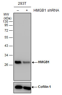
Non-transfected (–) and transfected (+) 293T whole cell extracts (30 µg) were separated by 12% SDS-PAGE, and the membrane was blotted with HMGB1 antibody [GT383] diluted at 1:2000. The HRP-conjugated anti-mouse IgG antibody was used to detect the primary antibody.Application
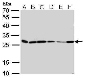
HMGB1 antibody [GT383] detects HMGB1 protein by western blot analysis. A. 30 µg Neuro2A whole cell lysate/extract B. 30 µg C8D30 whole cell lysate/extract C. 30 µg NIH-3T3 whole cell lysate/extract D. 30 µg BCL-1 whole cell lysate/extract E. 30 µg Raw264.7 whole cell lysate/extract F. 30 µg C2C12 whole cell lysate/extract 12% SDS-PAGE HMGB1 antibody [GT383] dilution: 1:500 The HRP-conjugated anti-mouse IgG antibody was used to detect the primary antibody.Application
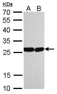
HMGB1 antibody [GT383] detects HMGB1 protein by western blot analysis. A. 30 µg PC-12 whole cell lysate/extract B. 30 µg Rat-2 whole cell lysate/extract 12% SDS-PAGE HMGB1 antibody [GT383] dilution: 1:1000 The HRP-conjugated anti-mouse IgG antibody was used to detect the primary antibody.Application
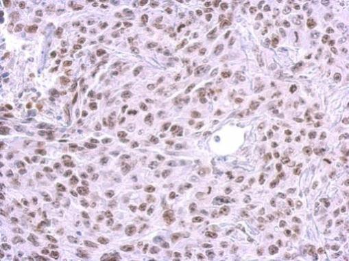
HMGB1 antibody [GT383] detects HMGB1 protein at nucleus on SAS xenograft by immunohistochemical analysis. Sample: Paraffin-embedded SAS xenograft. HMGB1 antibody [GT383] dilution: 1:200.Application
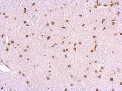
HMGB1 antibody [GT383] detects HMGB1 protein at nucleus on mouse brain by immunohistochemical analysis. Sample: Paraffin-embedded mouse brain. HMGB1 antibody [GT383] dilution: 1:200.Application
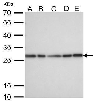
HMGB1 antibody [GT383] detects HMGB1 protein by western blot analysis. A. 30 µg 293T whole cell lysate/extract B. 30 µg A431 whole cell lysate/extract C. 30 µg HeLa whole cell lysate/extract D. 30 µg HepG2 whole cell lysate/extract E. 30 µg A375 whole cell lysate/extract 12% SDS-PAGE HMGB1 antibody [GT383] dilution: 1:1000 The HRP-conjugated anti-mouse IgG antibody was used to detect the primary antibody.| Product Name | HMGB1 antibody |
|---|---|
| Antibody Type | Primary Antibodies |
| Product description | Mouse Monoclonal antibody to HMGB1 (high-mobility group box 1)DNA binding proteins that associates with chromatin and has the ability to bend DNA. Binds preferentially single-stranded DNA. Involved in V(D)J recombination by acting as a cofactor of the RAG complex. Acts by stimulating cleavage and RAG protein binding at the 23 bp spacer of conserved recombination signal sequences (RSS). Heparin-binding protein that has a role in the extension of neurite-type cytoplasmic processes in developing cells. |
| Immunogen | Recombinant protein encompassing a sequence within the center region of human HMGB1. The exact sequence is proprietary. |
| Clonality | Monoclonal |
|---|---|
| Isotype | IgG2 |
| Clone Number | GT383 |
| Host Species | Mouse |
| Tested Applications | IHC-PWB |
| *Optimal dilutions/concentrations should be determined by the researcher.
: |
|
| Species Reactivity | HumanMouseRat |
| Predicted Reactivity | Xenopus laevis, Dog, Pig, Rabbit, Chicken, Rhesus Monkey, BovineXenopus laevis (93%), Dog (100%), Pig (100%), Rabbit (100%), Chicken (94%), Rhesus Monkey (100%), Bovine (100%) |
| Concentration | 1mg/ml |
| Purification | Affinity purified |
| Gene Synonyms | DKFZp686A04236 antibody HMG1 antibody HMG3 antibody SBP-1 antibody HMGB1 antibody HMG-1 antibody Amphoterin antibody high-mobility group box 1 antibody high mobility group protein B1 antibody high mobility group protein 1 antibody Sulfoglucuronyl |
|---|---|
| Molecular Weight(MW) | 25 kDa (note) (3) |
| Cellular Localization | Nucleus |
Application
Non-transfected (–) and transfected (+) 293T whole cell extracts (30 µg) were separated by 12% SDS-PAGE, and the membrane was blotted with HMGB1 antibody [GT383] diluted at 1:2000. The HRP-conjugated anti-mouse IgG antibody was used to detect the primary antibody.
Application
HMGB1 antibody [GT383] detects HMGB1 protein by western blot analysis. A. 30 µg Neuro2A whole cell lysate/extract B. 30 µg C8D30 whole cell lysate/extract C. 30 µg NIH-3T3 whole cell lysate/extract D. 30 µg BCL-1 whole cell lysate/extract E. 30 µg Raw264.7 whole cell lysate/extract F. 30 µg C2C12 whole cell lysate/extract 12% SDS-PAGE HMGB1 antibody [GT383] dilution: 1:500 The HRP-conjugated anti-mouse IgG antibody was used to detect the primary antibody.
Application
HMGB1 antibody [GT383] detects HMGB1 protein by western blot analysis. A. 30 µg PC-12 whole cell lysate/extract B. 30 µg Rat-2 whole cell lysate/extract 12% SDS-PAGE HMGB1 antibody [GT383] dilution: 1:1000 The HRP-conjugated anti-mouse IgG antibody was used to detect the primary antibody.
Application
HMGB1 antibody [GT383] detects HMGB1 protein at nucleus on SAS xenograft by immunohistochemical analysis. Sample: Paraffin-embedded SAS xenograft. HMGB1 antibody [GT383] dilution: 1:200.
Application
HMGB1 antibody [GT383] detects HMGB1 protein at nucleus on mouse brain by immunohistochemical analysis. Sample: Paraffin-embedded mouse brain. HMGB1 antibody [GT383] dilution: 1:200.
Application
HMGB1 antibody [GT383] detects HMGB1 protein by western blot analysis. A. 30 µg 293T whole cell lysate/extract B. 30 µg A431 whole cell lysate/extract C. 30 µg HeLa whole cell lysate/extract D. 30 µg HepG2 whole cell lysate/extract E. 30 µg A375 whole cell lysate/extract 12% SDS-PAGE HMGB1 antibody [GT383] dilution: 1:1000 The HRP-conjugated anti-mouse IgG antibody was used to detect the primary antibody.| Positive Control | 293T , A431 , HeLa , HepG2 , A375 , Neuro2A , C8D30 , NIH-3T3 , BCL-1 , Raw264.7 , C2C12 , PC-12 , Rat2 |
|---|---|
| Application Notes | *Optimal dilutions/concentrations should be determined by the researcher.
: |
| Form | Liquid |
|---|---|
| Storage Instructions | Keep as concentrated solution. Aliquot and store at -20ºC or below. Avoid multiple freeze-thaw cycles. |
| Storage Buffer | PBS |


