Application
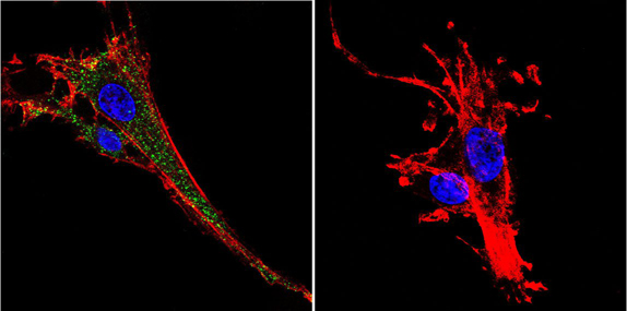
Immunofluorescent analysis of KCNQ2 using Anti-KCNQ2 Polyclonal Antibody shows staining in C6 Cells. KCNQ2 staining (green), F-Actin staining with Phalloidin (red) and nuclei with DAPI (blue) is shown. Cells were grown on chamber slides and fixed with formaldehyde prior to staining. Cells were probed without (control) or with or an antibody recognizing KCNQ2 at a dilution of 1:100 over night at 4 °C, washed with PBS and incubated with a DyLight-488 conjugated secondary antibody (Goat Anti-Rabbit). Images were taken at 60X magnification.Application
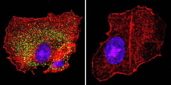
Immunofluorescent analysis of KCNQ2 using Anti-KCNQ2 Polyclonal Antibody shows staining in HepG2 Cells. KCNQ2 staining (green), F-Actin staining with Phalloidin (red) and nuclei with DAPI (blue) is shown. Cells were grown on chamber slides and fixed with formaldehyde prior to staining. Cells were probed without (control) or with or an antibody recognizing KCNQ2 at a dilution of 1:100 over night at 4 °C, washed with PBS and incubated with a DyLight-488 conjugated secondary antibody (Goat Anti-Rabbit). Images were taken at 60X magnification.Application
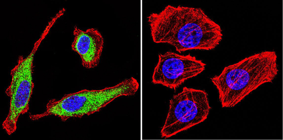
Immunofluorescent analysis of KCNQ2 using Anti-KCNQ2 Polyclonal Antibody shows staining in U251 Cells. KCNQ2 staining (green), F-Actin staining with Phalloidin (red) and nuclei with DAPI (blue) is shown. Cells were grown on chamber slides and fixed with formaldehyde prior to staining. Cells were probed without (control) or with or an antibody recognizing KCNQ2 at a dilution of 1:20 over night at 4 °C, washed with PBS and incubated with a DyLight-488 conjugated secondary antibody (Goat Anti-Rabbit). Images were taken at 60X magnification.| Product Name | KCNQ2 antibody |
|---|---|
| Antibody Type | Primary Antibodies |
| Product description | Rabbit Polyclonal antibody to KCNQ2KCNQs are members of the voltage-dependent non-inactivating potassium channel family. Currently there are five known KNCQs (KCNQ1-5) found in the central nervous system and KNCQ2 and 3 have demonstrated their importance in M-current activation. Studies have shown that KCNQ2 and KCNQ3 form heteromultimers that, when formed, substantially increase the M-current. Inhibition of M-current controls neuron excitability throughout the nervous system as well as the responsiveness to synaptic inputs. Genetic mutations in these proteins have been linked to disorders such as benign familial neonatal convulsions (BFNC), deafness, neuropathic pain and epilepsy. |
| Immunogen | GST fusion protein encoding the first 70 amino acids of human KCNQ2. |
| Clonality | Polyclonal |
|---|---|
| Host Species | Rabbit |
| Tested Applications | ICC/IFIHCIHC-FWB |
| ICC/IF: 1:500: |
|
| Species Reactivity | HumanMouseRat |
| Concentration | 1mg/ml (Please refer to the vial label for the specific concentration) |
| Purification | Affinity purified |
| Gene Synonyms | KCNQ2 BFNC BFNS1 EBN EBN1 EIEE7 ENB1 HNSPC KCNA11 KV7.2 KVEBN1 potassium voltage-gated channel KQT-like subfamily member 2 |
|---|---|
| Target | KCNQ2 |
Application
Immunofluorescent analysis of KCNQ2 using Anti-KCNQ2 Polyclonal Antibody shows staining in C6 Cells. KCNQ2 staining (green), F-Actin staining with Phalloidin (red) and nuclei with DAPI (blue) is shown. Cells were grown on chamber slides and fixed with formaldehyde prior to staining. Cells were probed without (control) or with or an antibody recognizing KCNQ2 at a dilution of 1:100 over night at 4 °C, washed with PBS and incubated with a DyLight-488 conjugated secondary antibody (Goat Anti-Rabbit). Images were taken at 60X magnification.
Application
Immunofluorescent analysis of KCNQ2 using Anti-KCNQ2 Polyclonal Antibody shows staining in HepG2 Cells. KCNQ2 staining (green), F-Actin staining with Phalloidin (red) and nuclei with DAPI (blue) is shown. Cells were grown on chamber slides and fixed with formaldehyde prior to staining. Cells were probed without (control) or with or an antibody recognizing KCNQ2 at a dilution of 1:100 over night at 4 °C, washed with PBS and incubated with a DyLight-488 conjugated secondary antibody (Goat Anti-Rabbit). Images were taken at 60X magnification.
Application
Immunofluorescent analysis of KCNQ2 using Anti-KCNQ2 Polyclonal Antibody shows staining in U251 Cells. KCNQ2 staining (green), F-Actin staining with Phalloidin (red) and nuclei with DAPI (blue) is shown. Cells were grown on chamber slides and fixed with formaldehyde prior to staining. Cells were probed without (control) or with or an antibody recognizing KCNQ2 at a dilution of 1:20 over night at 4 °C, washed with PBS and incubated with a DyLight-488 conjugated secondary antibody (Goat Anti-Rabbit). Images were taken at 60X magnification.| Application Notes | ICC/IF: 1:500: |
|---|
| Form | Liquid |
|---|---|
| Storage Instructions | Keep as concentrated solution. Aliquot and store at -20ºC or below. Avoid multiple freeze-thaw cycles. |
| Storage Buffer | (1 mg/ml) in PBS containing 1 mg/ml BSA, glycerol, and 0.02% sodium azide. |


