Application
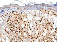
HT-29 xenograft tumor in mouse 20x pretreatment w/ citrate pH 6.0 using anti-Ki67 antibody at 5 ug/mL for 30 min at RT.Application
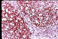
Immunohistochemical analysis of formalin-fixed paraffin-embedded human tonsil using Ki-67 antibody. Signal is detected using ABC method with AEC chromogenApplication
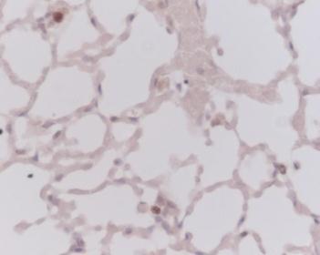
This picture was kindly supplied as part of the review submitted by Dr Robert O'Donogue. Tissue sections from mouse lung were stained with at a dilution of 1/50.| Product Name | Ki67 antibody |
|---|---|
| Antibody Type | Primary Antibodies |
| Product description | Rabbit polyclonal to Ki67-Proliferation MarkerKi-67 antigen is the prototypic cell cycle related nuclear protein, expressed by proliferating cells in all phases of the active cell cycle (G1, S, G2 and M phase). It is absent in resting (G0) cells. Ki67 antibodies are useful in establishing the cell growing fraction in neoplasms (immunohistochemically quantified by determining the number of Ki67-positive cells among the total number of resting cells = Ki67 index). In neoplastic tissues the prognostic value is comparable to the tritiated thymidine-labelling index. The correlation between low Ki67 index and histologically low-grade tumours is strong. |
| Immunogen | Synthetic peptide from the human Ki-67 allantigen. |
| Clonality | Polyclonal |
|---|---|
| Isotype | IgG |
| Host Species | Rabbit |
| Tested Applications | ICC/IFIHCIHC-P |
| For IHC-P: Use at a concentration of 1 0 - 2 0 μg / m l f o r 3 0 m i n a t R T. Staining of formalin/paraffin tissues requires boiling tissue sections in 10mM citrate buffer, pH 6.0, for 10-20 min followed by cooling at RT for 20 min. Optimal dilutions/concentrations should be determined by the end user. | |
| Species Reactivity | HumanMouseRat |
| Concentration | 1mg/ml (Please refer to the vial label for the specific concentration) |
| Purification | Affinity purified |
| Gene Synonyms | KIA |
|---|---|
| Target | Ki67 |
| Cellular Localization | Nuclear |
Application
HT-29 xenograft tumor in mouse 20x pretreatment w/ citrate pH 6.0 using anti-Ki67 antibody at 5 ug/mL for 30 min at RT.
Application
Immunohistochemical analysis of formalin-fixed paraffin-embedded human tonsil using Ki-67 antibody. Signal is detected using ABC method with AEC chromogen
Application
This picture was kindly supplied as part of the review submitted by Dr Robert O'Donogue. Tissue sections from mouse lung were stained with at a dilution of 1/50.| Application Notes | For IHC-P: Use at a concentration of 1 0 - 2 0 μg / m l f o r 3 0 m i n a t R T. Staining of formalin/paraffin tissues requires boiling tissue sections in 10mM citrate buffer, pH 6.0, for 10-20 min followed by cooling at RT for 20 min. Optimal dilutions/concentrations should be determined by the end user. |
|---|
| Form | Liquid |
|---|---|
| Storage Instructions | Keep as concentrated solution, aliquot and store at 4°C. Do not freeze. |
| Storage Buffer | 10mM Phosphate-buffered saline containing 0.2% BSA and 0.1% sodium azide |


