Application
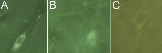
at a dilution of 1/1000, staining L1CAM (green; Alexa 488 secondary at 1/2000) on 30m coronal rat brain tissue sections in free floating IHC. Images showing neuron body, cytoplasm and axon labeling: [A] neuron; 40x objective [B] neuron and axons;| Product Name | L1CAM antibody |
|---|---|
| Antibody Type | Primary Antibodies |
| Product description | Mouse monoclonal [UJ127] to L1CAMThe L1CAM gene, which is located in Xq28, is involved in three distinct conditions: 1) HSAS (hydrocephalus-stenosis of the aqueduct of Sylvius); 2) MASA (mental retardation, aphasia, shuffling gait, adductus thumbs); and 3) SPG1 (spastic paraplegia). The L1, neural cell adhesion molecule (L1CAM) also plays an important role in axon growth, fasciculation, neural migration and in mediating neuronal differentiation. Expression of L1 protein is restricted to tissues arising from neuroectoderm. |
| Immunogen | Homogenous suspension of 16 week human fetal brain. |
| Clonality | Monoclonal |
|---|---|
| Isotype | IgG1 |
| Clone Number | UJ127 |
| Host Species | Mouse |
| Tested Applications | IHCIHC-PIPWB |
| Immunoprecipitation (natured and denatured): use at a concentration of 2μg / mg. Western Blotting: use at a concentration of 1 - 2μg / ml for 2hrs at RT. Immunohistochemistry (formalin fixed paraffin embedded): use at a concentration of 1 - 2μg / ml for 30 min at RT. Optimal dilutions/concentrations should be determined by the end user. By Western blot, this antibody detects a band of 220-240 kDa, which corresponds to the predicted molecular weight of L1CAM. Staining of formalin-fixed tissues requires boiling tissue sections in 10mM citrate buffer, pH 6.0, for 10-20 min followed by cooling at RT for 20 min.: |
|
| Species Reactivity | HumanRat |
| Concentration | 0.2mg/ml |
| Purification | Protein G |
| Gene Synonyms | CAML1 CD171 HSAS HSAS1 MASA MIC5 N-CAM-L1 N-CAML1 NCAM-L1 S10 SPG1 N CAML1 NCAM L1 N CAM L1 NCAML1 |
|---|---|
| Target | L1CAM |
| Tissue Specificity | is useful in the identification of primitive neuroectodermal tumors. It binds to tumors of neuroectodermal and glial origin. It does not bind to pediatric or adult brain. |
| Light Chain Type | kappa |
Application
at a dilution of 1/1000, staining L1CAM (green; Alexa 488 secondary at 1/2000) on 30m coronal rat brain tissue sections in free floating IHC. Images showing neuron body, cytoplasm and axon labeling: [A] neuron; 40x objective [B] neuron and axons;| Positive Control | IMR-5 cells |
|---|---|
| Application Notes | Immunoprecipitation (natured and denatured): use at a concentration of 2μg / mg. Western Blotting: use at a concentration of 1 - 2μg / ml for 2hrs at RT. Immunohistochemistry (formalin fixed paraffin embedded): use at a concentration of 1 - 2μg / ml for 30 min at RT. Optimal dilutions/concentrations should be determined by the end user. By Western blot, this antibody detects a band of 220-240 kDa, which corresponds to the predicted molecular weight of L1CAM. Staining of formalin-fixed tissues requires boiling tissue sections in 10mM citrate buffer, pH 6.0, for 10-20 min followed by cooling at RT for 20 min.: |
| Form | Liquid |
|---|---|
| Storage Instructions | Keep as concentrated solution, aliquot and store at 4°C. Do not freeze. |
| Storage Buffer | 10mM PBS, pH7.4, 0.2%BSA, 0.09% sodium azide |


