Application
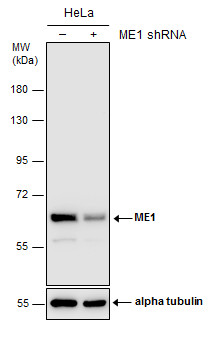
Non-transfected (–) and transfected (+) HeLa whole cell extracts (30 µg) were separated by 7.5% SDS-PAGE, and the membrane was blotted with ME1 antibody [GT15611] diluted at 1:1000.Application
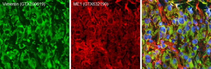
Double-labeled immunofluorescence photomicrographs of frozen sections of mouse brain. Green: Vimentin antibody diluted at 1:200. The signal was developed using goat anti-rabbit IgG antibody (Dylight488) . Red: ME1 antibody [GT15611] diluted at 1:200. The signal was developed using goat anti-mouse IgG antibody (Dylight594) . Blue: Nuclear staining with Hoechst 33342.Application
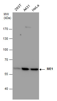
ME1 antibody detects ME1 protein by western blot analysis. Various whole cell extracts (30 μg) were separated by 7.5% SDS-PAGE, and the membrane was blotted with ME1 antibody diluted at a dilution of 1:1000.Application
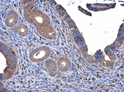
ME1 antibody [GT15611] detects ME1 protein at cytoplasm in mouse cervix by immunohistochemical analysis. Sample: Paraffin-embedded mouse cervix. ME1 antibody [GT15611] diluted at 1:500.Application
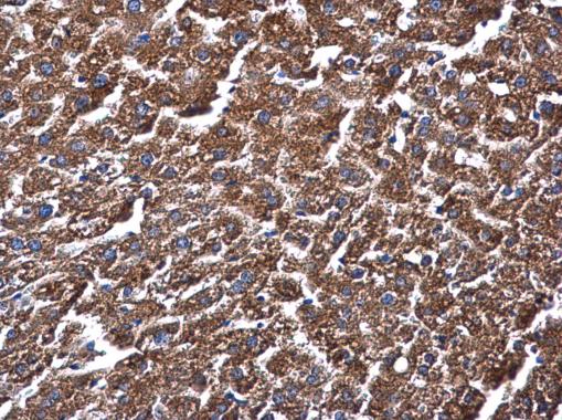
ME1 antibody [GT15611] detects ME1 protein at cytoplasm in mouse liver by immunohistochemical analysis. Sample: Paraffin-embedded mouse liver. ME1 antibody [GT15611] diluted at 1:500.| Product Name | ME1 antibody |
|---|---|
| Antibody Type | Primary Antibodies |
| Product description | Mouse Monoclonal antibody [GT15611] to ME1 (malic enzyme 1, NADP(+)-dependent, cytosolic)This gene encodes a cytosolic, NADP-dependent enzyme that generates NADPH for fatty acid biosynthesis. The activity of this enzyme, the reversible oxidative decarboxylation of malate, links the glycolytic and citric acid cycles. The regulation of expression for this gene is complex. Increased expression can result from elevated levels of thyroid hormones or by higher proportions of carbohydrates in the diet. [provided by RefSeq] |
| Immunogen | Recombinant protein encompassing a sequence within the center region of human ME1. The exact sequence is proprietary. |
| Clonality | Monoclonal |
|---|---|
| Isotype | IgG2a |
| Clone Number | GT15611 |
| Host Species | Mouse |
| Tested Applications | IHC-FIHC-PWB |
| *Optimal dilutions/concentrations should be determined by the researcher.
: |
|
| Species Reactivity | HumanMouse |
| Predicted Reactivity | Rat, Chicken, Sheep, BovineRat (91%), Chicken (80%), Sheep (92%), Bovine (91%) |
| Concentration | 1mg/ml (Please refer to the vial label for the specific concentration) |
| Purification | Affinity purified |
| Gene Synonyms | HUMNDME malic enzyme 1 NADP(+)-dependent cytosolic MES |
|---|---|
| Target | ME1 |
| Molecular Weight(MW) | 64 kDa (note) |
| Cellular Localization | Cytoplasm |
Application
Non-transfected (–) and transfected (+) HeLa whole cell extracts (30 µg) were separated by 7.5% SDS-PAGE, and the membrane was blotted with ME1 antibody [GT15611] diluted at 1:1000.
Application
Double-labeled immunofluorescence photomicrographs of frozen sections of mouse brain. Green: Vimentin antibody diluted at 1:200. The signal was developed using goat anti-rabbit IgG antibody (Dylight488) . Red: ME1 antibody [GT15611] diluted at 1:200. The signal was developed using goat anti-mouse IgG antibody (Dylight594) . Blue: Nuclear staining with Hoechst 33342.
Application
ME1 antibody detects ME1 protein by western blot analysis. Various whole cell extracts (30 μg) were separated by 7.5% SDS-PAGE, and the membrane was blotted with ME1 antibody diluted at a dilution of 1:1000.
Application
ME1 antibody [GT15611] detects ME1 protein at cytoplasm in mouse cervix by immunohistochemical analysis. Sample: Paraffin-embedded mouse cervix. ME1 antibody [GT15611] diluted at 1:500.
Application
ME1 antibody [GT15611] detects ME1 protein at cytoplasm in mouse liver by immunohistochemical analysis. Sample: Paraffin-embedded mouse liver. ME1 antibody [GT15611] diluted at 1:500.| Positive Control | 293T , A431 , HeLa |
|---|---|
| Application Notes | *Optimal dilutions/concentrations should be determined by the researcher.
: |
| Form | Liquid |
|---|---|
| Storage Instructions | Keep as concentrated solution. Aliquot and store at -20ºC or below. Avoid multiple freeze-thaw cycles. |
| Storage Buffer | PBS |


