Application
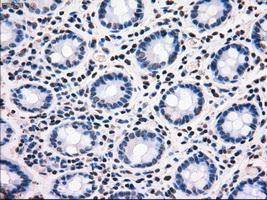
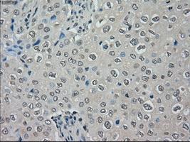
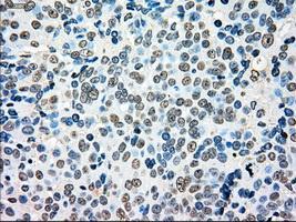
Immunohistochemical staining of paraffin-embedded colon tissue using anti-MRI1mouse monoclonal antibody. ( Dilution 1:50)Application
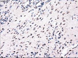
Immunohistochemical staining of paraffin-embedded Ovary tissue using anti-MRI1mouse monoclonal antibody. ( Dilution 1:50)Application
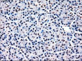
Immunohistochemical staining of paraffin-embedded pancreas tissue using anti-MRI1mouse monoclonal antibody. ( Dilution 1:50)Application
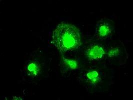
Anti-MRI1 mouse monoclonal antibody immunofluorescent staining of COS7 cells transiently transfected with MRI1Application
Immunohistochemical staining of paraffin-embedded Adenocarcinoma of breast tissue using anti-MRI1 mouse monoclonal antibody. ( Dilution 1:50)Application
Immunohistochemical staining of paraffin-embedded Adenocarcinoma of ovary tissue using anti-MRI1mouse monoclonal antibody. ( Dilution 1:50)Application
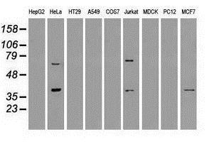
Western blot analysis of extracts (35ug) from 9 different cell lines by using anti-anti-MRI1monoclonal antibody.Application
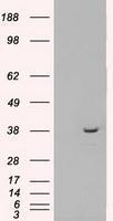
HEK293T cells were transfected with the pCMV6-ENTRY control (Left lane) or pCMV6-ENTRY MRI1 (Right lane) cDNA for 48 hrs and lysed. Equivalent amounts of cell lysates (5 ug per lane) were separated by SDS-PAGE and immunoblotted with anti-MRI1.| Product Name | MRI1 antibody [6G9] |
|---|---|
| Antibody Type | Primary Antibodies |
| Product description | Mouse monoclonal antibody [6G9] to MRI1Catalyzes the interconversion of methylthioribose-1-phosphate (MTR-1-P) into methylthioribulose-1-phosphate (MTRu-1-P). Independently from catalytic activity, promotes cell invasion in response to constitutive RhoA activation by promoting FAK tyrosine phosphorylation and stress fiber turnover. |
| Immunogen | Protein expressed in 293T cell transfected with human MRI1 expression vector |
| Clonality | Monoclonal |
|---|---|
| Isotype | IgG1 |
| Clone Number | 6G9 |
| Host Species | Mouse |
| Tested Applications | ICC/IFIHCIHC-PWB |
| Optimal working dilutions should be determined experimentally by the end user. | |
| Species Reactivity | Human |
| Concentration | 1mg/ml |
| Purification | Affinity purified |
| Gene Synonyms | Ypr118w, MRI1, MGC3207, MTNA |
|---|---|
| Target | MRI1 |
| Tissue Specificity | is specific for human MRI1. |
| Cellular Localization | Primarily nuclear, but cytoplasmic in cancer cells, with enrichment at leading edge of the plasma membrane in late stage tumor cells. |
Application
Immunohistochemical staining of paraffin-embedded colon tissue using anti-MRI1mouse monoclonal antibody. ( Dilution 1:50)
Application
Immunohistochemical staining of paraffin-embedded Ovary tissue using anti-MRI1mouse monoclonal antibody. ( Dilution 1:50)
Application
Immunohistochemical staining of paraffin-embedded pancreas tissue using anti-MRI1mouse monoclonal antibody. ( Dilution 1:50)
Application
Anti-MRI1 mouse monoclonal antibody immunofluorescent staining of COS7 cells transiently transfected with MRI1
Application
Immunohistochemical staining of paraffin-embedded Adenocarcinoma of breast tissue using anti-MRI1 mouse monoclonal antibody. ( Dilution 1:50)
Application
Immunohistochemical staining of paraffin-embedded Adenocarcinoma of ovary tissue using anti-MRI1mouse monoclonal antibody. ( Dilution 1:50)
Application
Western blot analysis of extracts (35ug) from 9 different cell lines by using anti-anti-MRI1monoclonal antibody.
Application
HEK293T cells were transfected with the pCMV6-ENTRY control (Left lane) or pCMV6-ENTRY MRI1 (Right lane) cDNA for 48 hrs and lysed. Equivalent amounts of cell lysates (5 ug per lane) were separated by SDS-PAGE and immunoblotted with anti-MRI1.| Application Notes | Optimal working dilutions should be determined experimentally by the end user. |
|---|
| Form | Liquid |
|---|---|
| Storage Instructions | For short-term storage, store at 4°C or aliquot into working amounts and store at -20°C. For long-term storage, store at -70°C (aliquotted). Avoid repeated freeze-thaw cycles. |
| Storage Buffer | PBS (PH 7.3) containing 1% BSA, 50% glycerol and 0.02% sodium azide. |


