Application
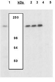
Extracts of Murine T cells were left untreated (1) or treated with PMA and Ca2+ ionophore ionomycin (2-5), were resolved by SDS-PAGE on a 10% Tris-glycine gel and transferred to PVDF. The membrane was blocked with a 5% BSA-TBST buffer overnight at 4 degree C, then incubated with the NFAT1 [pS54] (mouse) antibody in a 3% BSA-TBST buffer for two hours at room temperature, following prior incubation with: no peptide (1,2), the non-phosphopeptide corresponding to the phosphopeptide immunogen (3), a generic phosphoserine-containing peptide (4), or the phosphopeptide immunogen (5). After washing, the membrane was incubated with goat anti rabbit IgG alkaline phosphatase. The data show only the phopshopeptide corresponding to NFAT1 [pS54] (mouse) completely blocks the antibody signal, demonstrating specificity of the antibody. The data also show the upregulation of NFAT1 upon PMA Ca2+ ionophore ionomycin treatment in this cell system.| Product Name | NFAT1 (phospho Ser54) antibody |
|---|---|
| Antibody Type | Primary Antibodies |
| Product description | Rabbit polyclonal to NFAT1 ( phospho S54 )Nuclear factor of activated T cells (NFAT) is a family of transcription factors implicated in multiple biological processes including cytokine gene expression, cardiac hypertrophy and adipocyte differentiation. NFAT 1 (also known as NFATc2 or NFATp) is a 154 kDa member of this family that is regulated by the calcium-dependent phosphatase calcineurin. When calcineurin is activated by calcium it dephosphorylates multiple residues in the regulatory domain of NFAT 1, leading to its translocation to the nucleus and activation of its transcriptional activity. Once in the nucleus, NFAT proteins act synergistically with the AP-1 transcription factor complex to regulate the expression of multiple genes. Serine 54 in mouse NFAT 1 has been shown to be important in the regulation of its transcriptional activity. |
| Immunogen | Synthetic peptide - derived from a region of mouse NFAT 1 that contains serine 54 |
| Modification | p-Ser54 |
| Clonality | Polyclonal |
|---|---|
| Isotype | IgG |
| Host Species | Rabbit |
| Tested Applications | WB |
| For WB: Use at a dilution of 1:1000. Predicted molecular weight: 154 kDa. Optimal dilutions/concentrations should be determined by the end user.: |
|
| Species Reactivity | Mouse |
| Concentration | 1mg/ml |
| Purification | Affinity purified |
| Gene Synonyms | AI607462 NF-ATc2 NFAT1 NFAT1-D Nfatp NF ATc2 NFAT1 D NFATc2 NFAT1D NFAT1 (phospho S54) NFAT1 (phospho Ser54) NFAT1 (pS54) NFAT1 (pSer54) NFAT1 phospho S54 NFAT1 phospho Ser54 Phospho NFAT1 (S54) Phospho NFAT1 (Ser54) Phospho NFAT1 (pS54) |
|---|---|
| Target | NFAT1 |
Application
Extracts of Murine T cells were left untreated (1) or treated with PMA and Ca2+ ionophore ionomycin (2-5), were resolved by SDS-PAGE on a 10% Tris-glycine gel and transferred to PVDF. The membrane was blocked with a 5% BSA-TBST buffer overnight at 4 degree C, then incubated with the NFAT1 [pS54] (mouse) antibody in a 3% BSA-TBST buffer for two hours at room temperature, following prior incubation with: no peptide (1,2), the non-phosphopeptide corresponding to the phosphopeptide immunogen (3), a generic phosphoserine-containing peptide (4), or the phosphopeptide immunogen (5). After washing, the membrane was incubated with goat anti rabbit IgG alkaline phosphatase. The data show only the phopshopeptide corresponding to NFAT1 [pS54] (mouse) completely blocks the antibody signal, demonstrating specificity of the antibody. The data also show the upregulation of NFAT1 upon PMA Ca2+ ionophore ionomycin treatment in this cell system.| Positive Control | Murine T-cells + PMA + Ionomycin |
|---|---|
| Application Notes | For WB: Use at a dilution of 1:1000. Predicted molecular weight: 154 kDa. Optimal dilutions/concentrations should be determined by the end user.: |
| Form | Liquid |
|---|---|
| Storage Instructions | Keep as concentrated solution. Store at 4ºC short term. For extended storage aliquot and store at -20ºC or below. Avoid freeze-thaw cycles. |
| Storage Buffer | Phosphate-buffered saline, pH 7.3 containing 50% glycerol, 1.0 mg/ml BSA and 0.05% sodium azide |


