Application
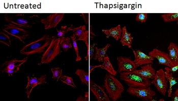
Immunofluorescent analysis of NFATc2 (green) in HeLa cells. Formalin fixed cells were permeabilized with 0.1% Triton X-100 in TBS for 10 minutes at room temperature and blocked with 1% Blocker BSA for 15 minutes at room temperature. Cells were left untreated (left panel) or treated with 1uM staurosporine (right panel) for 3 hours and probed with a NFATc2 monoclonal antibody , at a dilution of 1:100 for at least 1 hour at room temperature, washed with PBS, and incubated with DyLight 488 goat anti-mouse IgG secondary antibody at a dilution of 1:400 for 30 minutes at room temperature. F-Actin (red) was stained with DyLight 554 Phalloidin and nuclei (blue) were stained with Hoechst 33342 dye.Application
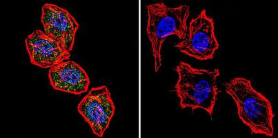
Immunofluorescent analysis of NFATc2 using NFATc2 Monoclonal Antibody shows staining in Hela Cells. NFATc2 (green), F-Actin staining with Phalloidin (red) and nuclei with DAPI (blue) is shown. Cells were grown on chamber slides and fixed with formaldehyde prior to staining. Cells were probed without (control) or with an antibody recognizing NFATc2 at a dilution of 1:20 overnight at 4 degree C, washed with PBS and incubated with a DyLight-488 conjugated secondary antibody.Application
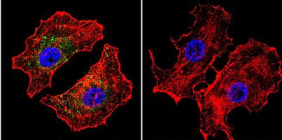
Immunofluorescent analysis of NFATc2 using NFATc2 monoclonal antibody shows staining in MCF-7 Cells. NFATc2 (green), F-Actin staining with Phalloidin (red) and nuclei with DAPI (blue) is shown. Cells were grown on chamber slides and fixed with formaldehyde prior to staining. Cells were probed without (control) or with an antibody recognizing NFATc2 at a dilution of 1:20 over night at 4 C, washed with PBS and incubated with a DyLight-488 conjugated secondary antibody.Application
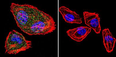
Immunofluorescent analysis of NFATc2 using NFATc2 monoclonal antibody shows staining in U251 Cells. NFATc2 (green), F-Actin staining with Phalloidin (red) and nuclei with DAPI (blue) is shown. Cells were grown on chamber slides and fixed with formaldehyde prior to staining. Cells were probed without (control) or with an antibody recognizing NFATc2 at a dilution of 1:20 over night at 4 degree C, washed with PBS and incubated with a DyLight-488 conjugated secondary antibody.Application
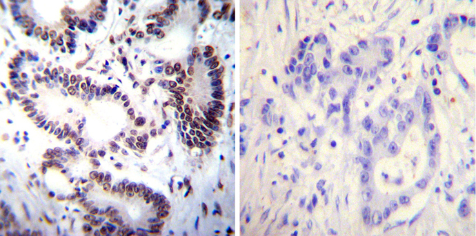
Immunohistochemistry was performed on cancer biopsies of deparaffinized Human colon carcinoma tissues. To expose target proteins, heat induced antigen retrieval was performed using 10mM sodium citrate (pH6.0) buffer, microwaved for 8-15 minutes. Following antigen retrieval tissues were blocked in 3% BSA-PBS for 30 minutes at room temperature. Tissues were then probed at a dilution of 1:100 with a mouse monoclonal antibody recognizing NFATc2 (MA1-025) or without primary antibody (negative control) overnight at 4°C in a humidified chamber. Tissues were washed extensively with PBST and endogenous peroxidase activity was quenched with a peroxidase suppressor. Detection was performed using a biotin-conjugated secondary antibody and SA-HRP, followed by colorimetric detection using DAB. Tissues were counterstained with hematoxylin and prepped for mounting.Application
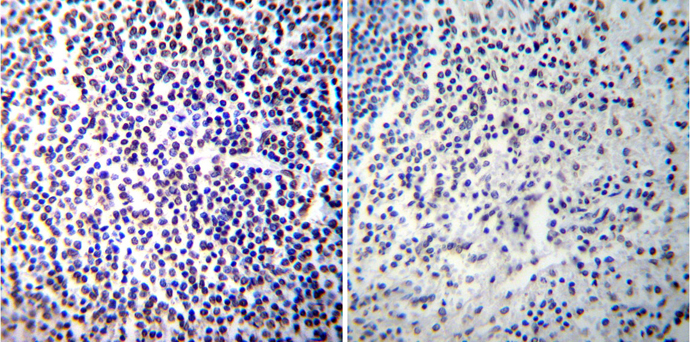
Immunohistochemistry was performed on normal de-paraffinized human spleen tissue tissues. To expose target proteins, heat induced antigen retrieval was performed using 10mM sodium citrate (pH6.0) buffer, microwaved for 8-15 minutes. Following antigen retrieval tissues were blocked in 3% BSA-PBS for 30 minutes at room temperature. Tissues were then probed at a dilution of 1:100 with a mouse monoclonal antibody recognizing NFATc2 or without primary antibody (negative control) overnight at 4°C in a humidified chamber. Tissues were washed extensively with PBST and endogenous peroxidase activity was quenched with a peroxidase suppressor. Detection was performed using a biotin-conjugated secondary antibody and SA-HRP, followed by colorimetric detection using DAB. Tissues were counterstained with hematoxylin and prepped for mounting.Application
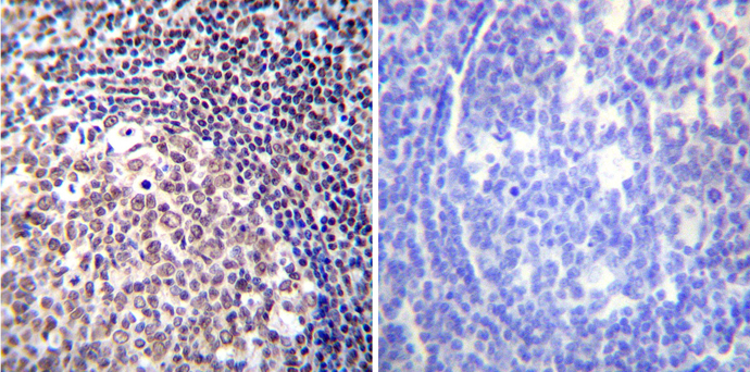
Immunohistochemistry was performed on normal de-paraffinized human tonsil tissue tissues. To expose target proteins, heat induced antigen retrieval was performed using 10mM sodium citrate (pH6.0) buffer, microwaved for 8-15 minutes. Following antigen retrieval tissues were blocked in 3% BSA-PBS for 30 minutes at room temperature. Tissues were then probed at a dilution of 1:100 with a mouse monoclonal antibody recognizing NFATc2 or without primary antibody (negative control) overnight at 4°C in a humidified chamber. Tissues were washed extensively with PBST and endogenous peroxidase activity was quenched with a peroxidase suppressor. Detection was performed using a biotin-conjugated secondary antibody and SA-HRP, followed by colorimetric detection using DAB. Tissues were counterstained with hematoxylin and prepped for mounting.| Product Name | NFAT1 antibody |
|---|---|
| Antibody Type | Primary Antibodies |
| Product description | Mouse monoclonal [25A10.D6.D2] to NFAT1The nuclear factor of activated T-cells (NFAT) transcription complex is required for the expression of a group of proteins that collectively regulate the immune response. Four NFAT proteins, encoded on separate genes and expressed as several splice variants, have been described: NFAT1 (also known as NFATp or NFATc2), NFAT2 (NFATc or NFATc1), NFAT3, and NFAT4 (NFATx or NFATc3). These proteins show a low level of sequence similarity with the Dorsal/Rel/NFkB family of transcription factors. Another NFAT-related protein termed NFAT5 differs from isoforms 1-4 in that it lacks many of the Fos/Jun contact sites observed in its predecessors and its subcellular localization is not calcineurin-dependent. |
| Immunogen | Synthetic peptide: (51)AISSPSGLAYPDDVLDYGL(69). The immunizing peptide corresponds to amino acid residues 51-69 of mouse NFAT1-A, B and C isoforms. The sequence of this N-terminal peptide is ~70% homologous with human NFAT1. |
| Clonality | Monoclonal |
|---|---|
| Isotype | IgG1 |
| Clone Number | 25A10.D6.D2 |
| Host Species | Mouse |
| Tested Applications | FACSGSAICC/IFIHCIHC-FIHC-PIPWBChIP |
| WB:1:500~1:1000 IP:1:100~1:500 ICC/IF:1:100~1:500 IHC:1:50~1:100 |
|
| Species Reactivity | HumanMouseRat |
| Concentration | 1mg/ml (Please refer to the vial label for the specific concentration) |
| Gene Synonyms | NFAT1 NFATP |
|---|---|
| Target | NFAT1 |
| Tissue Specificity | This antibody does not cross react with NFAT2 (NFATc, NFATc1). |
Application
Immunofluorescent analysis of NFATc2 (green) in HeLa cells. Formalin fixed cells were permeabilized with 0.1% Triton X-100 in TBS for 10 minutes at room temperature and blocked with 1% Blocker BSA for 15 minutes at room temperature. Cells were left untreated (left panel) or treated with 1uM staurosporine (right panel) for 3 hours and probed with a NFATc2 monoclonal antibody , at a dilution of 1:100 for at least 1 hour at room temperature, washed with PBS, and incubated with DyLight 488 goat anti-mouse IgG secondary antibody at a dilution of 1:400 for 30 minutes at room temperature. F-Actin (red) was stained with DyLight 554 Phalloidin and nuclei (blue) were stained with Hoechst 33342 dye.
Application
Immunofluorescent analysis of NFATc2 using NFATc2 Monoclonal Antibody shows staining in Hela Cells. NFATc2 (green), F-Actin staining with Phalloidin (red) and nuclei with DAPI (blue) is shown. Cells were grown on chamber slides and fixed with formaldehyde prior to staining. Cells were probed without (control) or with an antibody recognizing NFATc2 at a dilution of 1:20 overnight at 4 degree C, washed with PBS and incubated with a DyLight-488 conjugated secondary antibody.
Application
Immunofluorescent analysis of NFATc2 using NFATc2 monoclonal antibody shows staining in MCF-7 Cells. NFATc2 (green), F-Actin staining with Phalloidin (red) and nuclei with DAPI (blue) is shown. Cells were grown on chamber slides and fixed with formaldehyde prior to staining. Cells were probed without (control) or with an antibody recognizing NFATc2 at a dilution of 1:20 over night at 4 C, washed with PBS and incubated with a DyLight-488 conjugated secondary antibody.
Application
Immunofluorescent analysis of NFATc2 using NFATc2 monoclonal antibody shows staining in U251 Cells. NFATc2 (green), F-Actin staining with Phalloidin (red) and nuclei with DAPI (blue) is shown. Cells were grown on chamber slides and fixed with formaldehyde prior to staining. Cells were probed without (control) or with an antibody recognizing NFATc2 at a dilution of 1:20 over night at 4 degree C, washed with PBS and incubated with a DyLight-488 conjugated secondary antibody.
Application
Immunohistochemistry was performed on cancer biopsies of deparaffinized Human colon carcinoma tissues. To expose target proteins, heat induced antigen retrieval was performed using 10mM sodium citrate (pH6.0) buffer, microwaved for 8-15 minutes. Following antigen retrieval tissues were blocked in 3% BSA-PBS for 30 minutes at room temperature. Tissues were then probed at a dilution of 1:100 with a mouse monoclonal antibody recognizing NFATc2 (MA1-025) or without primary antibody (negative control) overnight at 4°C in a humidified chamber. Tissues were washed extensively with PBST and endogenous peroxidase activity was quenched with a peroxidase suppressor. Detection was performed using a biotin-conjugated secondary antibody and SA-HRP, followed by colorimetric detection using DAB. Tissues were counterstained with hematoxylin and prepped for mounting.
Application
Immunohistochemistry was performed on normal de-paraffinized human spleen tissue tissues. To expose target proteins, heat induced antigen retrieval was performed using 10mM sodium citrate (pH6.0) buffer, microwaved for 8-15 minutes. Following antigen retrieval tissues were blocked in 3% BSA-PBS for 30 minutes at room temperature. Tissues were then probed at a dilution of 1:100 with a mouse monoclonal antibody recognizing NFATc2 or without primary antibody (negative control) overnight at 4°C in a humidified chamber. Tissues were washed extensively with PBST and endogenous peroxidase activity was quenched with a peroxidase suppressor. Detection was performed using a biotin-conjugated secondary antibody and SA-HRP, followed by colorimetric detection using DAB. Tissues were counterstained with hematoxylin and prepped for mounting.
Application
Immunohistochemistry was performed on normal de-paraffinized human tonsil tissue tissues. To expose target proteins, heat induced antigen retrieval was performed using 10mM sodium citrate (pH6.0) buffer, microwaved for 8-15 minutes. Following antigen retrieval tissues were blocked in 3% BSA-PBS for 30 minutes at room temperature. Tissues were then probed at a dilution of 1:100 with a mouse monoclonal antibody recognizing NFATc2 or without primary antibody (negative control) overnight at 4°C in a humidified chamber. Tissues were washed extensively with PBST and endogenous peroxidase activity was quenched with a peroxidase suppressor. Detection was performed using a biotin-conjugated secondary antibody and SA-HRP, followed by colorimetric detection using DAB. Tissues were counterstained with hematoxylin and prepped for mounting.| Application Notes | WB:1:500~1:1000 IP:1:100~1:500 ICC/IF:1:100~1:500 IHC:1:50~1:100 |
|---|
| Form | Liquid |
|---|---|
| Storage Instructions | Keep as concentrated solution. Store at 4ºC short term. For extended storage aliquot and store at -20ºC or below. Avoid freeze-thaw cycles. |
| Storage Buffer | Phosphate-buffered saline containing 0.05% sodium azide |


