Application
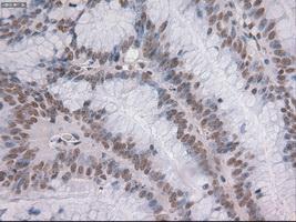
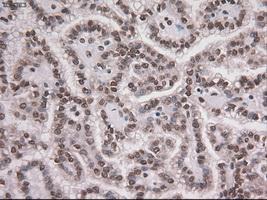
Immunohistochemical staining of paraffin-embedded Adenocarcinoma of colon tissue using anti-NOTCH1mouse monoclonal antibody. ( Dilution 1:50)Application
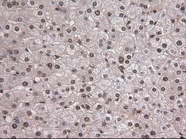
Immunohistochemical staining of paraffin-embedded liver tissue using anti-NOTCH1mouse monoclonal antibody. ( Dilution 1:50)Application
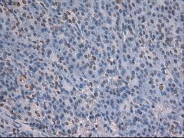
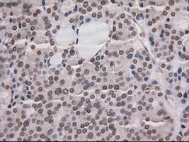
Immunohistochemical staining of paraffin-embedded pancreas tissue using anti-NOTCH1mouse monoclonal antibody. ( Dilution 1:50)Application
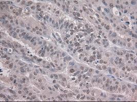
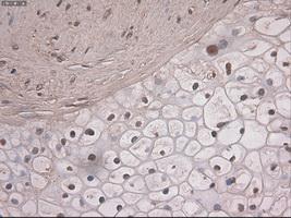
Immunohistochemical staining of paraffin-embedded Adenocarcinoma of ovary tissue using anti-NOTCH1mouse monoclonal antibody. ( Dilution 1:50)Application
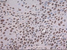
Immunohistochemical staining of paraffin-embedded Carcinoma of pancreas tissue using anti-NOTCH1mouse monoclonal antibody. ( Dilution 1:50)Application
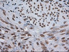
Immunohistochemical staining of paraffin-embedded Carcinoma of thyroid tissue using anti-NOTCH1mouse monoclonal antibody. ( Dilution 1:50)Application
Immunohistochemical staining of paraffin-embedded endometrium tissue using anti-NOTCH1mouse monoclonal antibody. ( Dilution 1:50)Application
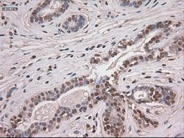
Immunohistochemical staining of paraffin-embedded breast tissue using anti-NOTCH1 mouse monoclonal antibody. ( Dilution 1:50)Application
Immunohistochemical staining of paraffin-embedded Carcinoma of kidney tissue using anti-NOTCH1mouse monoclonal antibody. ( Dilution 1:50)Application
Immunohistochemical staining of paraffin-embedded Carcinoma of lung tissue using anti-NOTCH1mouse monoclonal antibody. ( Dilution 1:50)Application
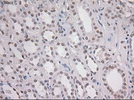
Immunohistochemical staining of paraffin-embedded Kidney tissue using anti-NOTCH1mouse monoclonal antibody. ( Dilution 1:50)Application
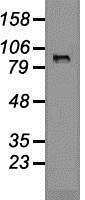
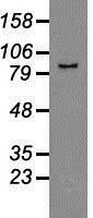
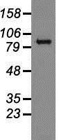
Western blot analysis of 35ug of cell extracts from human colon adenocarcinoma (HT29) cells using anti-NOTCH1 antibody.Application
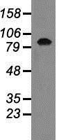
Western blot analysis of 35ug of cell extracts from human Lung adenocarcinoma (A549) cells using anti-NOTCH1 antibody.Application
Western blot analysis of 35ug of cell extracts from human Kidney (HEK293T) cells using anti-NOTCH1 antibody.Application
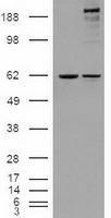
HEK293T cells were transfected with the pCMV6-ENTRY control (Left lane) or pCMV6-ENTRY NOTCH1 (Right lane) cDNA for 48 hrs and lysed. Equivalent amounts of cell lysates (5 ug per lane) were separated by SDS-PAGE and immunoblotted with anti-NOTCH1.Application
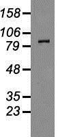
Western blot analysis of 35ug of cell extracts from human Liver carcinoma (HepG2) cells using anti-NOTCH1 antibody.Application
Western blot analysis of 35ug of cell extracts from human (HeLa) cells using anti-NOTCH1 antibody.| Product Name | NOTCH1 antibody [4C9] |
|---|---|
| Antibody Type | Primary Antibodies |
| Product description | Mouse monoclonal antibody [4C9] to Notch1Functions as a receptor for membrane-bound ligands Jagged1, Jagged2 and Delta1 to regulate cell-fate determination. Upon ligand activation through the released notch intracellular domain (NICD) it forms a transcriptional activator complex with RBP-J kappa and activates genes of the enhancer of split locus. Affects the implementation of differentiation, proliferation and apoptotic programs. May be important for normal lymphocyte function. In altered form, may contribute to transformation or progression in some T-cell neoplasms. Involved in the maturation of both CD4+ and CD8+ cells in the thymus. May be important for follicular differentiation and possibly cell fate selection within the follicle. During cerebellar development, may function as a receptor for neuronal DNER and may be involved in the differentiation of Bergmann glia. |
| Immunogen | Recombinant protein expressed in E.coli corresponding to amino acids 2300-2556 of human NOTCH1 |
| Clonality | Monoclonal |
|---|---|
| Isotype | IgG2 |
| Clone Number | 4C9 |
| Host Species | Mouse |
| Tested Applications | IHCIHC-PWB |
| Optimal working dilutions should be determined experimentally by the end user. | |
| Species Reactivity | Human |
| Concentration | 1mg/ml |
| Purification | Affinity purified |
| Gene Synonyms | NOTCH1, hN1, TAN1 |
|---|---|
| Target | Notch1 |
| Tissue Specificity | is specific for amino acids 2300-2556 of human NOTCH1 |
| Cellular Localization | Following proteolytical processing NICD is translocated to the nucleus. |
Application
Immunohistochemical staining of paraffin-embedded Adenocarcinoma of colon tissue using anti-NOTCH1mouse monoclonal antibody. ( Dilution 1:50)
Application
Immunohistochemical staining of paraffin-embedded liver tissue using anti-NOTCH1mouse monoclonal antibody. ( Dilution 1:50)
Application
Immunohistochemical staining of paraffin-embedded pancreas tissue using anti-NOTCH1mouse monoclonal antibody. ( Dilution 1:50)
Application
Immunohistochemical staining of paraffin-embedded Adenocarcinoma of ovary tissue using anti-NOTCH1mouse monoclonal antibody. ( Dilution 1:50)
Application
Immunohistochemical staining of paraffin-embedded Carcinoma of pancreas tissue using anti-NOTCH1mouse monoclonal antibody. ( Dilution 1:50)
Application
Immunohistochemical staining of paraffin-embedded Carcinoma of thyroid tissue using anti-NOTCH1mouse monoclonal antibody. ( Dilution 1:50)
Application
Immunohistochemical staining of paraffin-embedded endometrium tissue using anti-NOTCH1mouse monoclonal antibody. ( Dilution 1:50)
Application
Immunohistochemical staining of paraffin-embedded breast tissue using anti-NOTCH1 mouse monoclonal antibody. ( Dilution 1:50)
Application
Immunohistochemical staining of paraffin-embedded Carcinoma of kidney tissue using anti-NOTCH1mouse monoclonal antibody. ( Dilution 1:50)
Application
Immunohistochemical staining of paraffin-embedded Carcinoma of lung tissue using anti-NOTCH1mouse monoclonal antibody. ( Dilution 1:50)
Application
Immunohistochemical staining of paraffin-embedded Kidney tissue using anti-NOTCH1mouse monoclonal antibody. ( Dilution 1:50)
Application
Western blot analysis of 35ug of cell extracts from human colon adenocarcinoma (HT29) cells using anti-NOTCH1 antibody.
Application
Western blot analysis of 35ug of cell extracts from human Lung adenocarcinoma (A549) cells using anti-NOTCH1 antibody.
Application
Western blot analysis of 35ug of cell extracts from human Kidney (HEK293T) cells using anti-NOTCH1 antibody.
Application
HEK293T cells were transfected with the pCMV6-ENTRY control (Left lane) or pCMV6-ENTRY NOTCH1 (Right lane) cDNA for 48 hrs and lysed. Equivalent amounts of cell lysates (5 ug per lane) were separated by SDS-PAGE and immunoblotted with anti-NOTCH1.
Application
Western blot analysis of 35ug of cell extracts from human Liver carcinoma (HepG2) cells using anti-NOTCH1 antibody.
Application
Western blot analysis of 35ug of cell extracts from human (HeLa) cells using anti-NOTCH1 antibody.| Application Notes | Optimal working dilutions should be determined experimentally by the end user. |
|---|
| Form | Liquid |
|---|---|
| Storage Instructions | For short-term storage, store at 4°C or aliquot into working amounts and store at -20°C. For long-term storage, store at -70°C (aliquotted). Avoid repeated freeze-thaw cycles. |
| Storage Buffer | PBS (PH 7.3) containing 1% BSA, 50% glycerol and 0.02% sodium azide. |


