Application
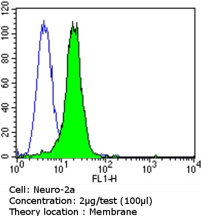
Flow cytometry analysis of PSD95 in Neuro-2a cells compared to an isotype control (blue). Cells were harvested, adjusted to a concentration of 1-5x10^6 cells/ml, fixed with 2% paraformaldehyde and washed with PBS. Cells were blocked with a 2% solution of BSA-PBS for 30 min at room temperature and incubated with PSD95 antibody [6G6-1C9] at a dilution of 2 ug/test for 60 min at room temperature. Cells were then incubated for 40 min at room temperature in the dark using a proper secondary antibody and re-suspended in PBS for FACS analysis.Application
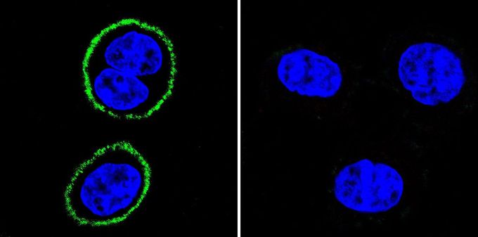
Immunofluorescent analysis of PSD95 in HeLa cells. PSD95 staining (green), F-Actin staining with Phalloidin (red) and nuclei with DAPI (blue) is shown. Cells were grown on slides and fixed with formaldehyde prior to staining. Cells were probed without (control) or with PSD95 antibody [6G6-1C9] at a dilution of 1:100-1:200 over night at 4 °C, washed with PBS and incubated with a proper secondary antibody. Images were taken at 60X magnification.Application
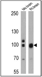
Western blot analysis of PSD95 in 25 ug of mouse brain and rat brain lysates. Proteins were transferred to a PVDF membrane and blocked at 4oC overnight. The membrane was probed with PSD95 antibody [6G6-1C9] at a dilution of 1:200 overnight at 4°C, washed in TBST, and probed with an HRP-conjugated secondary antibody for 1 hr at room temperature in the dark. Chemiluminescent detection was performed.| Product Name | PSD95 antibody [6G6-1C9] |
|---|---|
| Antibody Type | Primary Antibodies |
| Product description | Mouse monoclonal antibody [6G6-1C9] to PSD95a molecular scaffolding protein that binds and clusters N-methyl-D-aspartate receptors at neuronal synapses; may be involved in guanine nucleotide-mediated signal transduction pathway [RGD, Feb 2006] |
| Immunogen | Purified recombinant rat PSD-95. |
| Clonality | Monoclonal |
|---|---|
| Isotype | IgG2a |
| Clone Number | 6G6-1C9 |
| Host Species | Mouse |
| Tested Applications | BlockingELISAFACSICC/IFIHCIPWBChIP |
| ICC/IF: 1:50 - 1:2000 | |
| Species Reactivity | HumanMouseRatPrimate |
| Concentration | 1mg/ml |
| Purification | Protein G |
| Gene Synonyms | discs, large homolog 4 (Drosophila) , Dlgh4 , Dlg4 , Sap90 |
|---|---|
| Target | PSD95 |
Application
Flow cytometry analysis of PSD95 in Neuro-2a cells compared to an isotype control (blue). Cells were harvested, adjusted to a concentration of 1-5x10^6 cells/ml, fixed with 2% paraformaldehyde and washed with PBS. Cells were blocked with a 2% solution of BSA-PBS for 30 min at room temperature and incubated with PSD95 antibody [6G6-1C9] at a dilution of 2 ug/test for 60 min at room temperature. Cells were then incubated for 40 min at room temperature in the dark using a proper secondary antibody and re-suspended in PBS for FACS analysis.
Application
Immunofluorescent analysis of PSD95 in HeLa cells. PSD95 staining (green), F-Actin staining with Phalloidin (red) and nuclei with DAPI (blue) is shown. Cells were grown on slides and fixed with formaldehyde prior to staining. Cells were probed without (control) or with PSD95 antibody [6G6-1C9] at a dilution of 1:100-1:200 over night at 4 °C, washed with PBS and incubated with a proper secondary antibody. Images were taken at 60X magnification.
Application
Western blot analysis of PSD95 in 25 ug of mouse brain and rat brain lysates. Proteins were transferred to a PVDF membrane and blocked at 4oC overnight. The membrane was probed with PSD95 antibody [6G6-1C9] at a dilution of 1:200 overnight at 4°C, washed in TBST, and probed with an HRP-conjugated secondary antibody for 1 hr at room temperature in the dark. Chemiluminescent detection was performed.| Application Notes | ICC/IF: 1:50 - 1:2000 |
|---|
| Form | Liquid |
|---|---|
| Storage Instructions | -20° C, Avoid Freeze/Thaw Cycles |
| Storage Buffer | PBS containing 0.05 % sodium azide |


