Application
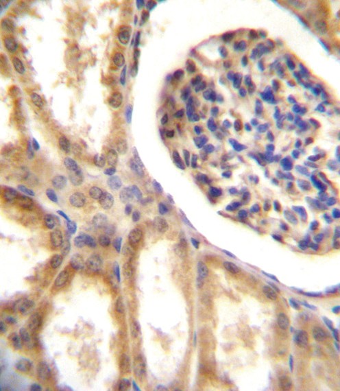
DDR1 Antibody immunohistochemistry analysis in formalin fixed and paraffin embedded human kidney tissue followed by peroxidase conjugation of the secondary antibody and DAB staining.Application
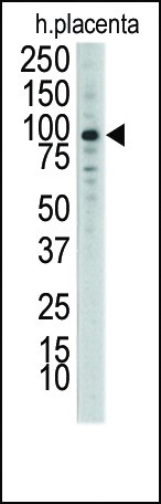
Western blot analysis of anti-DDR1 Antibody Pab in placenta lysateApplication
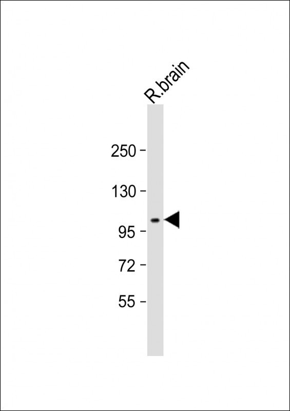
Western Blot at 1:2000 dilution + rat brain lysate Lysates/proteins at 20 ug per lane.Application
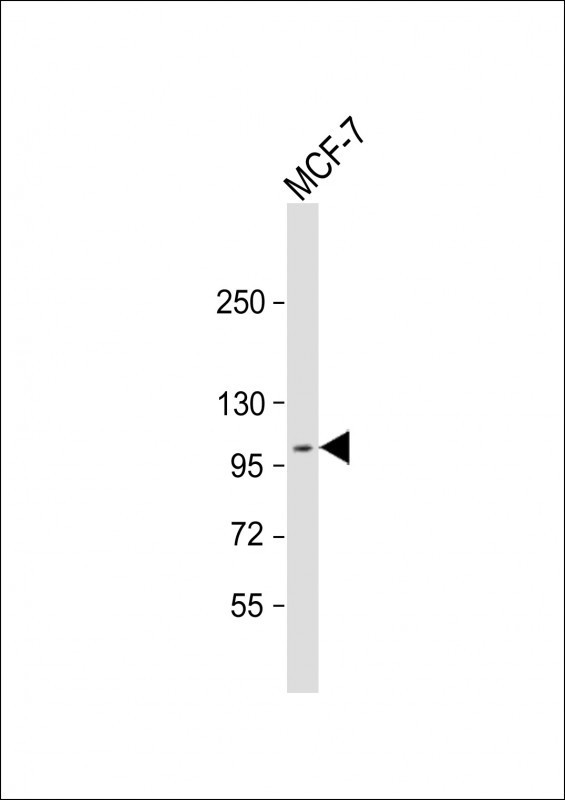
Western Blot at 1:2000 dilution + MCF-7 whole cell lysate Lysates/proteins at 20 ug per lane.Application
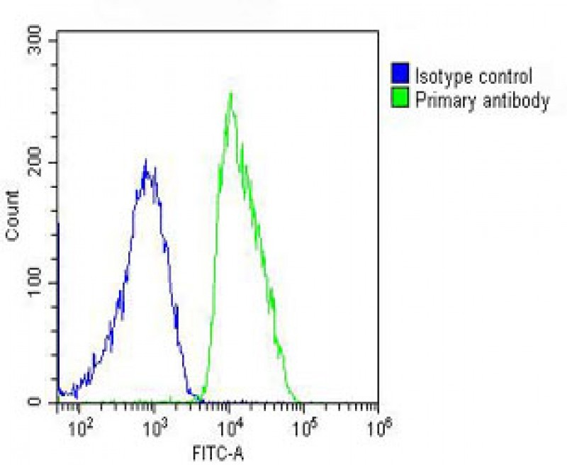
Overlay histogram showing MCF-7 cells stained with Antibody (green line). The cells were fixed with 2% paraformaldehyde (10 min) and then permeabilized with 90% methanol for 10 min. The cells were then icubated in 2% bovine serum albumin to block non-specific protein-protein interactions followed by the antibody (1:25 dilution) for 60 min at 37ºC. The secondary antibody used was Goat-Anti-Rabbit I| Product Name | DDR1 Antibody |
|---|---|
| Antibody Type | Primary Antibodies |
| Product description | Receptor tyrosine kinases (RTKs) play a key role in the communication of cells with their microenvironment. These molecules are involved in the regulation of cell growth, differentiation and metabolism. MCK10 is a RTK that is widely expressed in normal and transformed epithelial cells and is activated by various types of collagen. This protein belongs to a subfamily of tyrosine kinase receptors with a homology region to the Dictyostelium discoideum protein discoidin I in their extracellular domain. Its autophosphorylation is achieved by all collagens so far tested (type I to type VI). In situ studies and Northern-blot analysis showed that expression of this encoded protein is restricted to epithelial cells, particularly in the kidney, lung, gastrointestinal tract, and brain. In addition, this protein is significantly over-expressed in several human tumors from breast, ovarian, esophageal, and pediatric brain. The gene is located on chromosome 6p21.3 in proximity to several HLA class I genes.1) Vogel, W., et al., Mol. Cell 1(1):13-23 (1997). Playford, M.P., et al., Genome Res. 6(7):620-627 (1996). Perez, J.L., et al., Oncogene 12(7):1469-1477 (1996). Valent, A., et al., Hum. Genet. 98(1):12-15 (1996). Edelhoff, S., et al., Genomics 25(1):309-311 (1995). |
| Immunogen | This DDR1 antibody is generated from rabbits immunized with a KLH conjugated synthetic peptide between 17-47 amino acids from the N-terminal region of human DDR1. |
| Clonality | Polyclonal |
|---|---|
| Isotype | Ig |
| Host Species | Rabbit |
| Tested Applications | FACSIHC-PWB |
| For FACS starting dilution is: 1:25 For WB starting dilution is: 1:2000 For IHC-P starting dilution is: 1:10~50 | |
| Species Reactivity | HumanRat |
| Concentration | 1mg/ml |
| Purification | Affinity purified |
| Gene Symbol | DDR1 |
|---|---|
| Alternative Names | Epithelial discoidin domain-containing receptor 1 Epithelial discoidin domain receptor 1 CD167 antigen-like family member A Cell adhesion kinase Discoidin receptor tyrosine kinase HGK2 Mammary carcinoma kinase 10 MCK-10 Protein-tyrosine kinase 3A |
| Molecular Weight(MW) | 101 kDa |
| Sequence Similarities | Predicted species reactivity based on immunogen sequence: Mouse |
Application
DDR1 Antibody immunohistochemistry analysis in formalin fixed and paraffin embedded human kidney tissue followed by peroxidase conjugation of the secondary antibody and DAB staining.
Application
Western blot analysis of anti-DDR1 Antibody Pab in placenta lysate
Application
Western Blot at 1:2000 dilution + rat brain lysate Lysates/proteins at 20 ug per lane.
Application
Western Blot at 1:2000 dilution + MCF-7 whole cell lysate Lysates/proteins at 20 ug per lane.
Application
Overlay histogram showing MCF-7 cells stained with Antibody (green line). The cells were fixed with 2% paraformaldehyde (10 min) and then permeabilized with 90% methanol for 10 min. The cells were then icubated in 2% bovine serum albumin to block non-specific protein-protein interactions followed by the antibody (1:25 dilution) for 60 min at 37ºC. The secondary antibody used was Goat-Anti-Rabbit I| Application Notes | For FACS starting dilution is: 1:25 For WB starting dilution is: 1:2000 For IHC-P starting dilution is: 1:10~50 |
|---|
| Form | Liquid |
|---|---|
| Storage Instructions | Store at 4˚C for three months and -20˚C, stable for up to one year. As with all antibodies care should be taken to avoid repeated freeze thaw cycles. Antibodies should not be exposed to prolonged high temperatures. |
| Storage Buffer | Supplied in PBS with 0.09% (W/V) sodium azide. |


