Application
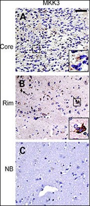
IHC of matched glioma core (top row) and rim (middle row) samples as well as normal brain control (bottom row) from a glioma invasion specific tissue microarray. Insets, individual tumor cells (black squares) are presented at higher magnification to facilitate interpretation of the staining pattern. Staining for MKK3 and pMKK3 exhibits strong signal in majority of invasive cells (B ) compared withApplication
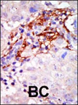
Formalin-fixed and paraffin-embedded human cancer tissue reacted with the primary antibody, which was peroxidase-conjugated to the secondary antibody, followed by AEC staining. BC = breast carcinoma; HC = hepatocarcinoma.Application
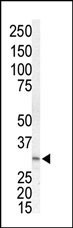
Western blot analysis of anti-MKK3 Pab in Jurkat cell lysate| Product Name | MEK3 Antibody |
|---|---|
| Antibody Type | Primary Antibodies |
| Product description | MAP2K3 is a dual specificity protein kinase that belongs to the MAP kinase kinase family. This kinase is activated by mitogenic and environmental stress, and participates in the MAP kinase-mediated signaling cascade. It phosphorylates and thus activates MAPK14/p38-MAPK. This kinase can be activated by insulin, and is necessary for the expression of glucose transporter. Expression of RAS oncogene is found to result in the accumulation of the active form of this kinase, which thus leads to the constitutive activation of MAPK14, and confers oncogenic transformation of primary cells. The inhibition of this kinase is involved in the pathogenesis of Yersina pseudotuberculosis.1) Yustein, J.T., et al., Oncogene 22(40):6129-6141 (2003). |
| Immunogen | This MEK3 (MAP2K3) antibody is generated from rabbits immunized with a KLH conjugated synthetic peptide between 15-45 amino acids from the N-terminal region of human MEK3 (MAP2K3). |
| Clonality | Polyclonal |
|---|---|
| Isotype | Ig |
| Host Species | Rabbit |
| Tested Applications | IHC-P,WB |
| For WB starting dilution is: 1:1000 For IHC-P starting dilution is: 1:50~100 | |
| Species Reactivity | Human |
| Concentration | 1mg/ml |
| Purification | Unpurified |
| Gene Symbol | MAP2K3 |
|---|---|
| Alternative Names | Dual specificity mitogen-activated protein kinase kinase 3 MAP kinase kinase 3 MAPKK 3 MAPK/ERK kinase 3 MEK 3 Stress-activated protein kinase kinase 2 SAPK kinase 2 SAPKK-2 SAPKK2 MAP2K3 MEK3 MKK3 PRKMK3 SKK2 |
| Molecular Weight(MW) | 39 kDa |
Application
IHC of matched glioma core (top row) and rim (middle row) samples as well as normal brain control (bottom row) from a glioma invasion specific tissue microarray. Insets, individual tumor cells (black squares) are presented at higher magnification to facilitate interpretation of the staining pattern. Staining for MKK3 and pMKK3 exhibits strong signal in majority of invasive cells (B ) compared with
Application
Formalin-fixed and paraffin-embedded human cancer tissue reacted with the primary antibody, which was peroxidase-conjugated to the secondary antibody, followed by AEC staining. BC = breast carcinoma; HC = hepatocarcinoma.
Application
Western blot analysis of anti-MKK3 Pab in Jurkat cell lysate| Application Notes | For WB starting dilution is: 1:1000 For IHC-P starting dilution is: 1:50~100 |
|---|
| Form | Liquid |
|---|---|
| Storage Instructions | Store at 4˚C for three months and -20˚C, stable for up to one year. As with all antibodies care should be taken to avoid repeated freeze thaw cycles. Antibodies should not be exposed to prolonged high temperatures. |
| Storage Buffer | Supplied in PBS with 0.09% (W/V) sodium azide. |


