Application
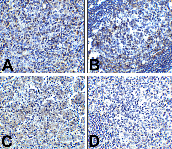
Immunohistochemistry of PD-1 in (A) human tonsil tissue, (B) human lymph node tissue, and (C) human spleen tissue with PD-1 antibody at 5 μg/mL. (D) Immunohistochemistry in human tonsil tissue with control mouse IgG staining at 5 μg/mL.Application
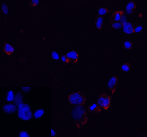
Immunofluorescence of PD-1 in transfected 293 cells with PD-1 antibody at 5 μg/mL. Lower left: Immunofluorescence in transfected 293 cells with control mouse IgG antibody at 5 μg/mL.Application
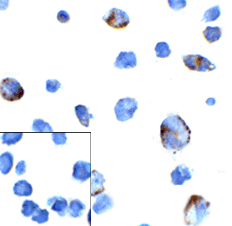
Immunocytochemistry of PD-1 in transfected 293 cells with PD-1 antibody at 5 μg/mL. Lower left: Immunocytochemistry in transfected 293 cells with control mouse IgG antibody at 5 μg/mL.Application
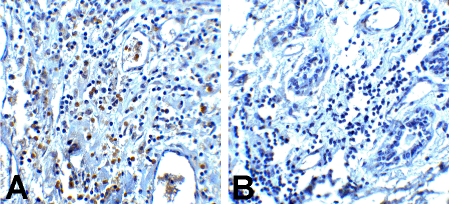
Immunohistochemistry of PD-1 in (A) human breast cancer tissue and (B) human normal breast tissue with PD-1 antibody at 5 μg/mL.Application
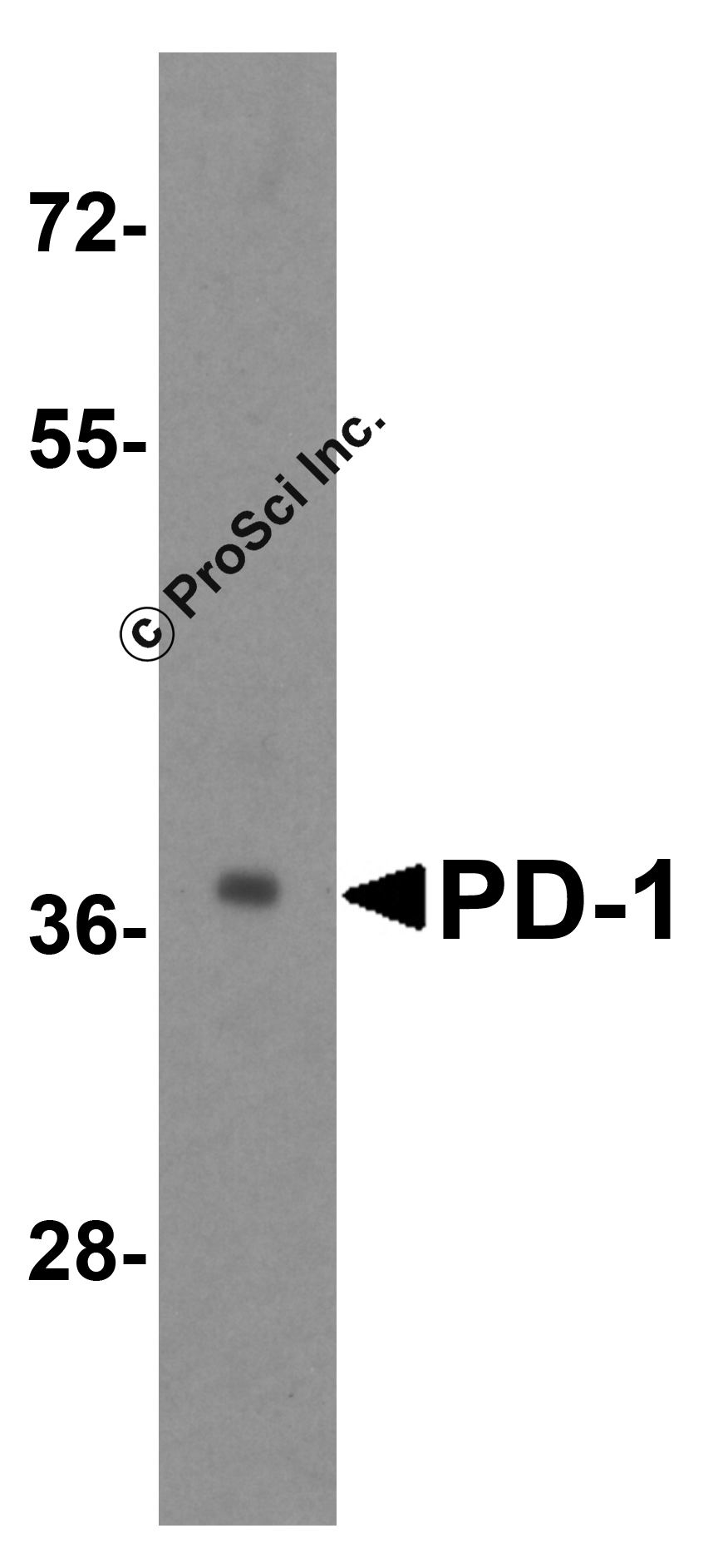
Western blot analysis of PD-1 in transfected 293 cell lysate with PD-1 antibody at 1 μg/mL.| Product Name | PD-1 Antibody |
|---|---|
| Antibody Type | Primary Antibodies |
| Antigen Alias | PD-1 Antibody: PD1, PD-1, CD279, SLEB2, hPD-1, hPD-l, hSLE1, PD1, Programmed cell death protein 1, Protein PD-1, PDCD1, PDCD-1 |
| Product description | Cell-mediated immune responses are initiated by T lymphocytes that are themselves stimulated by cognate peptides bound to MHC molecules on antigen-presenting cells (APC). T-cell activation is generally self-limited as activated T cells express receptors such as PD-1 (also known as PDCD-1) that mediate inhibitory signals from the APC. PD-1 can bind two different but related ligands, PDL-1 and PDL-2. Upon binding to either of these ligands, signals generated by PD-1 inhibit the activation of the immune response in the absence of "danger signals" such as LPS or other molecules associated with bacteria or other pathogens. Evidence for this is seen in PD-1-null mice who exhibit hyperactivated immune systems and autoimmune diseases. PD-1 is thus one of a growing number of immune checkpoint proteins.1) Holling TM, Schooten E, and van Den Elsing PJ. Function and regulation of MHC class II molecules in T-lymphocytes: of mice and men. Hum. Immunol. 2004; 65:282-90. |
| Immunogen | PD-1 antibody was raised against the extracellular domain of human PD-1. |
| Clonality | Monoclonal |
|---|---|
| Isotype | IgG1 |
| Host Species | Mouse |
| Tested Applications | ELISAICCIFIHC-PWB |
| PD-1 antibody can be used for detection of PD-1 by Western blot at 1 μg/mL. Antibody can also be used for immunohistochemistry starting at 5 μg/mL. For immunofluorescence start at 20 μg/mL.: |
|
| Species Reactivity | Human |
| Concentration | 1mg/ml |
| Purification | Protein A |
| Gene Symbol | PDCD1 |
|---|---|
| Alternative Names | PD-1 Antibody: PD1 PD-1 CD279 SLEB2 hPD-1 hPD-l hSLE1 PD1 Programmed cell death protein 1 Protein PD-1 PDCD1 PDCD-1 |
| Molecular Weight(MW) | Predicted: 32 kDa Observed: 38 kDa |
| Sequence Similarities | 3) Cat. No. 1369 - Human Lymph Node Tissue Lysate |
Application
Immunohistochemistry of PD-1 in (A) human tonsil tissue, (B) human lymph node tissue, and (C) human spleen tissue with PD-1 antibody at 5 μg/mL. (D) Immunohistochemistry in human tonsil tissue with control mouse IgG staining at 5 μg/mL.
Application
Immunofluorescence of PD-1 in transfected 293 cells with PD-1 antibody at 5 μg/mL. Lower left: Immunofluorescence in transfected 293 cells with control mouse IgG antibody at 5 μg/mL.
Application
Immunocytochemistry of PD-1 in transfected 293 cells with PD-1 antibody at 5 μg/mL. Lower left: Immunocytochemistry in transfected 293 cells with control mouse IgG antibody at 5 μg/mL.
Application
Immunohistochemistry of PD-1 in (A) human breast cancer tissue and (B) human normal breast tissue with PD-1 antibody at 5 μg/mL.
Application
Western blot analysis of PD-1 in transfected 293 cell lysate with PD-1 antibody at 1 μg/mL.| Positive Control | 1) Cat. No. 1306 - Human Spleen Tissue Lysate |
|---|---|
| Application Notes | PD-1 antibody can be used for detection of PD-1 by Western blot at 1 μg/mL. Antibody can also be used for immunohistochemistry starting at 5 μg/mL. For immunofluorescence start at 20 μg/mL.: |
| Form | Liquid |
|---|---|
| Storage Instructions | PD-1 antibody can be stored at 4˚C for three months and -20˚C, stable for up to one year. As with all antibodies care should be taken to avoid repeated freeze thaw cycles. Antibodies should not be exposed to prolonged high temperatures. |
| Storage Buffer | PD-1 Antibody is supplied in PBS containing 0.02% sodium azide. |


