WB
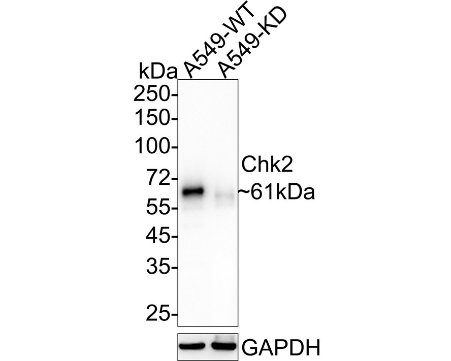
Western blot analysis of Chk2 on different lysates with Rabbit anti-Chk2 antibody at 1/1,000 dilution. Lane 1: A549-si NT cell lysate, Lane 2: A549-si Chk2 cell lysate, Lysates/proteins at 10 µg/Lane. Exposure time: 40 seconds; 4-20% SDS-PAGE gel. Proteins were transferred to a PVDF membrane and blocked with 5% NFDM/TBST for 1 hour at room temperature. The primary antibody at 1/1,000 dilution was used in 5% NFDM/TBST at 4℃ overnight. Goat Anti-Rabbit IgG - HRP Secondary Antibody at 1/50,000 dilution was used for 1 hour at room temperature.IHC
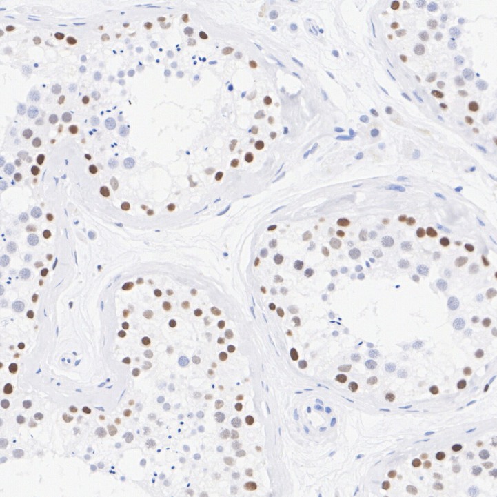
Immunohistochemical analysis of paraffin-embedded human testis tissue with Rabbit anti-Chk2 antibody at 1/1,000 dilution. The section was pre-treated using heat mediated antigen retrieval with sodium citrate buffer (pH 6.0) for 2 minutes. The tissues were blocked in 1% BSA for 20 minutes at room temperature, washed with ddH2O and PBS, and then probed with the primary antibody at 1/1,000 dilution for 1 hour at room temperature. The detection was performed using an HRP conjugated compact polymer system. DAB was used as the chromogen. Tissues were counterstained with hematoxylin and mounted with DPX.ICC/IF
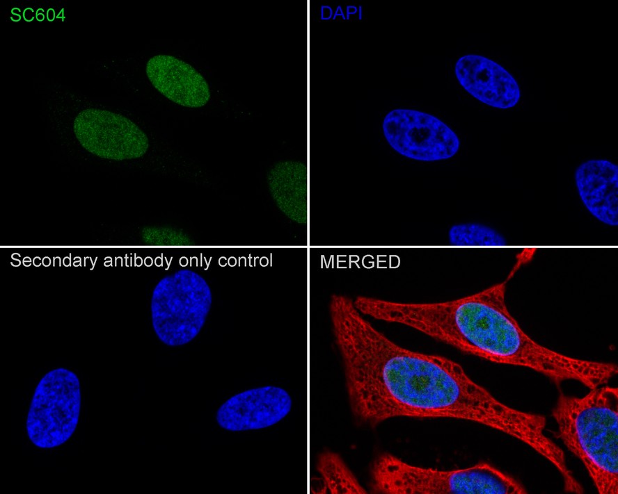
Immunocytochemistry analysis of HeLa cells labeling Chk2 with Rabbit anti-Chk2 antibody at 1/50 dilution. Cells were fixed in 4% paraformaldehyde for 15 minutes at room temperature, permeabilized with 0.1% Triton X-100 in PBS for 15 minutes at room temperature, then blocked with 1% BSA in 10% negative goat serum for 1 hour at room temperature. Cells were then incubated with Rabbit anti-Chk2 antibody at 1/50 dilution in 1% BSA in PBST overnight at 4 ℃. Goat Anti-Rabbit IgG H&L (488) was used as the secondary antibody at 1/1,000 dilution. PBS instead of the primary antibody was used as the secondary antibody only control. Nuclear DNA was labelled in blue with DAPI. Beta tubulin (red) was stained at 1/100 dilution overnight at +4℃. Goat Anti-Mouse IgG H&L (594) was used as the secondary antibody at 1/1,000 dilution.IP
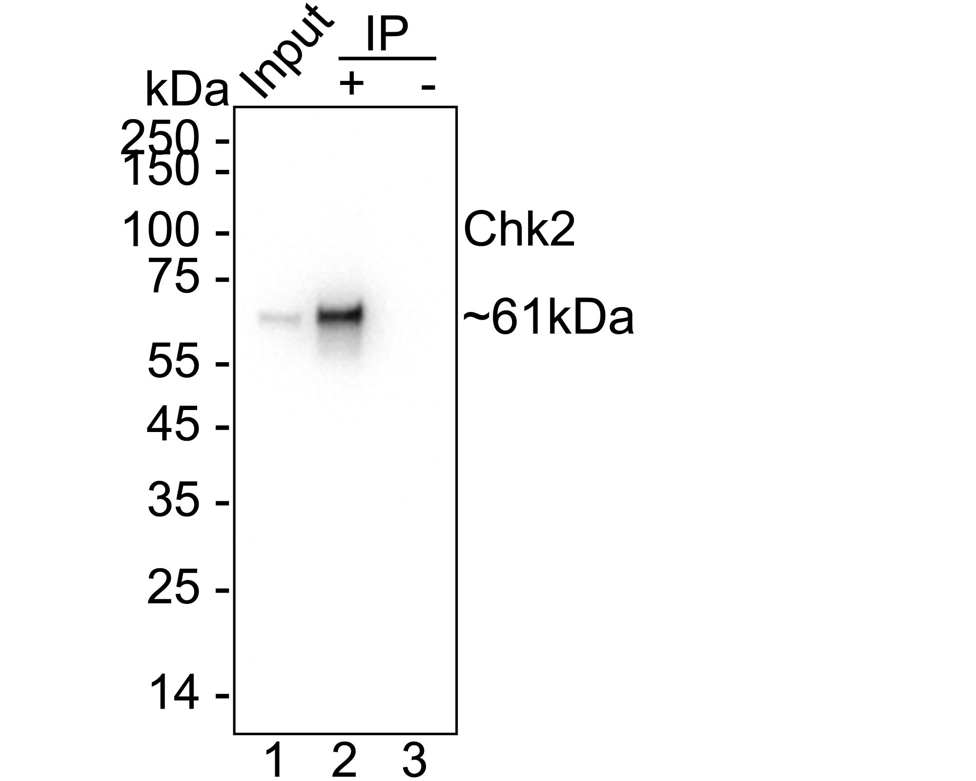
Chk2 was immunoprecipitated from 0.2 mg A549 cell lysate with Rabbit anti-Chk2 antibody at 2 µg/10 µl beads. Western blot was performed from the immunoprecipitate using Rabbit anti-Chk2 antibody at 1/2,000 dilution. HRP Conjugated Anti-Rabbit IgG for IP Nano-secondary antibody at 1/5,000 dilution was used for 1 hour at room temperature. Lane 1: A549 cell lysate (input), Lane 2: Rabbit anti-Chk2 antibody IP in A549 cell lysate, Lane 3: Rabbit IgG instead of Rabbit anti-Chk2 antibody in A549 cell lysate, Blocking/Dilution buffer: 5% NFDM/TBST Exposure time: 10 seconds.| Product Name | Chk2 Recombinant Rabbit Monoclonal Antibody |
|---|---|
| Antibody Type | Primary Antibodies |
| Product description | Cell cycle events are regulated by the sequential activation and deactivation of cyclin dependent kinases (Cdks) and by proteolysis of cyclins. Chk1 and Chk2 are involved in these processes as regulators of Cdks. Chk1 and Chk2 both function as essential components in the G2 DNA damage checkpoint by phosphorylating Cdc25C in response to DNA damage. Phosphorylation inhibits Cdc25C activity, thereby blocking mitosis. Cdc25A, Cdc25B and Cdc25C protein tyrosine phosphatases function as mitotic activators by dephosphorylating Cdc2 p34 on regulatory tyrosine residues. It has also been shown that Chk1 can phosphorylate Wee1 in vitro, providing evidence that the hyperphosphorylated form of Wee1, seen in cells delayed by Chk1 overexpression, is due to phosphorylation by Chk1. |
| Immunogen | Recombinant protein within Human Chk2 aa 10-200 / 543. |
| Clonality | monoclonal |
|---|---|
| Isotype | IgG |
| Host Species | Recombinant rabbit |
| Tested Applications | ICC/IFIHCIPWB |
| WB:1:1000-1:2000 IHC:1:1000 ICC/IF:1:50 IP:1-2μg/sample |
|
| Species Reactivity | Human |
| Concentration | 1mg/ml |
| Purification | Protein A |
| Gene Symbol | CHEK2 |
|---|---|
| Gene Synonyms | CDS1 CHK2 LFS2 RAD53 TPDS4 hCds1 HuCds1 PP1425 |
| Gene Full Name | checkpoint kinase 2 |
| Gene Summary | In response to DNA damage and replication blocks, cell cycle progression is halted through the control of critical cell cycle regulators. The protein encoded by this gene is a cell cycle checkpoint regulator and putative tumor suppressor. It contains a forkhead-associated protein interaction domain essential for activation in response to DNA damage and is rapidly phosphorylated in response to replication blocks and DNA damage. When activated, the encoded protein is known to inhibit CDC25C phosphatase, preventing entry into mitosis, and has been shown to stabilize the tumor suppressor protein p53, leading to cell cycle arrest in G1. In addition, this protein interacts with and phosphorylates BRCA1, allowing BRCA1 to restore survival after DNA damage. Mutations in this gene have been linked with Li-Fraumeni syndrome, a highly penetrant familial cancer phenotype usually associated with inherited mutations in TP53. Also, mutations in this gene are thought to confer a predisposition to sarcomas, breast cancer, and brain tumors. This nuclear protein is a member of the CDS1 subfamily of serine/threonine protein kinases. Several transcript variants encoding different isoforms have been found for this gene. [provided by RefSeq, Apr 2012] |
| Alternative Names | CDS 1 antibody Cds1 antibody Cds1 homolog antibody Checkpoint kinase 2 antibody Checkpoint like protein CHK2 antibody CHEK 2 antibody Chek2 antibody Chk 2 antibody CHK2 checkpoint homolog (S. pombe) antibody CHK2 checkpoint homolog antibody CHK2_HUMAN antibody hCds1 antibody HuCds 1 antibody LFS 2 antibody LFS2 antibody PP1425 antibody RAD 53 antibody RAD53 antibody Rad53 homolog antibody Serine/threonine protein kinase Chk2 antibody Serine/threonine-protein kinase Chk2 antibody |
| Molecular Weight(MW) | 61kDa |
| Cellular Localization | Nucleus. |
| SwissProt ID | O96017 |
|---|
WB
Western blot analysis of Chk2 on different lysates with Rabbit anti-Chk2 antibody at 1/1,000 dilution. Lane 1: A549-si NT cell lysate, Lane 2: A549-si Chk2 cell lysate, Lysates/proteins at 10 µg/Lane. Exposure time: 40 seconds; 4-20% SDS-PAGE gel. Proteins were transferred to a PVDF membrane and blocked with 5% NFDM/TBST for 1 hour at room temperature. The primary antibody at 1/1,000 dilution was used in 5% NFDM/TBST at 4℃ overnight. Goat Anti-Rabbit IgG - HRP Secondary Antibody at 1/50,000 dilution was used for 1 hour at room temperature.
IHC
Immunohistochemical analysis of paraffin-embedded human testis tissue with Rabbit anti-Chk2 antibody at 1/1,000 dilution. The section was pre-treated using heat mediated antigen retrieval with sodium citrate buffer (pH 6.0) for 2 minutes. The tissues were blocked in 1% BSA for 20 minutes at room temperature, washed with ddH2O and PBS, and then probed with the primary antibody at 1/1,000 dilution for 1 hour at room temperature. The detection was performed using an HRP conjugated compact polymer system. DAB was used as the chromogen. Tissues were counterstained with hematoxylin and mounted with DPX.
ICC/IF
Immunocytochemistry analysis of HeLa cells labeling Chk2 with Rabbit anti-Chk2 antibody at 1/50 dilution. Cells were fixed in 4% paraformaldehyde for 15 minutes at room temperature, permeabilized with 0.1% Triton X-100 in PBS for 15 minutes at room temperature, then blocked with 1% BSA in 10% negative goat serum for 1 hour at room temperature. Cells were then incubated with Rabbit anti-Chk2 antibody at 1/50 dilution in 1% BSA in PBST overnight at 4 ℃. Goat Anti-Rabbit IgG H&L (488) was used as the secondary antibody at 1/1,000 dilution. PBS instead of the primary antibody was used as the secondary antibody only control. Nuclear DNA was labelled in blue with DAPI. Beta tubulin (red) was stained at 1/100 dilution overnight at +4℃. Goat Anti-Mouse IgG H&L (594) was used as the secondary antibody at 1/1,000 dilution.
IP
Chk2 was immunoprecipitated from 0.2 mg A549 cell lysate with Rabbit anti-Chk2 antibody at 2 µg/10 µl beads. Western blot was performed from the immunoprecipitate using Rabbit anti-Chk2 antibody at 1/2,000 dilution. HRP Conjugated Anti-Rabbit IgG for IP Nano-secondary antibody at 1/5,000 dilution was used for 1 hour at room temperature. Lane 1: A549 cell lysate (input), Lane 2: Rabbit anti-Chk2 antibody IP in A549 cell lysate, Lane 3: Rabbit IgG instead of Rabbit anti-Chk2 antibody in A549 cell lysate, Blocking/Dilution buffer: 5% NFDM/TBST Exposure time: 10 seconds.| Positive Control | MCF-7, Hela, 293, human spleen tissue, human breast carcinoma tissue. |
|---|---|
| Application Notes | WB:1:1000-1:2000 IHC:1:1000 ICC/IF:1:50 IP:1-2μg/sample |
| Form | Liquid |
|---|---|
| Storage Instructions | Store at +4℃ after thawing. Aliquot store at -20℃ or -80℃. Avoid repeated freeze / thaw cycles. |
| Storage Buffer | 1*TBS (pH7.4), 1%BSA, 40%Glycerol. Preservative: 0.05% Sodium Azide. |


