



WB
Western blot analysis using CHK2 antibody against HepG2 (1) cell lysate.12% SDS-PAGE gel.Sample loading: 20μg /lane. Transfer the proteins onto a PVDF membrane (OM790003), and block it with TBST (OM750016) plus skimmed milk powder(OM750084) for one hour. Dilute the primary antibody with the antibody diluent (OM750012) at a ratio of 1:1000, and incubate it overnight at 4°C. Wash the membrane three times with TBST (OM750016), 5 minutes each time. At room temperature, dilute the secondary antibody, Goat Anti-Mouse IgG (H&L) - HRP(OM644366), at a ratio of 1:20000 and incubate for one hour. Wash the membrane three times with TBST (OM750016) again, 5 minutes each time. Use ECL (OM625701) for luminescence.staining time: 60S.IHC
Immunohistochemical analysis of paraffin-embedded Human prostate cancer tissues using CHK2 antibody with DAB staining.Pre-treat the sections with heat-mediated antigen retrieval using sodium citrate buffer (pH 6.0) (OM750020) for 2 minutes. Wash the sections with ddH₂O and PBS (OM750003). Block the tissue with 10% non-immune goat serum(OM760028) at room temperature for 30 minutes. Incubate the tissue with the primary antibody diluted at a ratio of 1:1500 at 4°C overnight. At room temperature, dilute the secondary antibody, Goat Anti-Mouse IgG (H&L) - HRP(OM644366), at a ratio of 1:200 and incubate for one hour. Use DAB(OM760029)as the chromogenic agent. Counterstain the tissue with hematoxylin, and mount the tissue sections with neutral gum.ICC/IF
Immunofluorescence analysis of HepG2 cells using CHK2 antibody (green).Cells are fixed in 4% paraformaldehyde at room temperature for 20 minutes. Then, they are permeabilized with a PBS (OM750003) solution containing 0.1% Triton X-100(OM750021) at room temperature for 15 minutes. Subsequently, the cells are blocked with 10% non - immune goat serum(OM760028) at room temperature for 1 hour.The cells are incubated overnight at 4°C with the primary antibody diluted 1:100 in PBS. The secondary antibody, Omnimabs® 488 Goat Anti-Mouse IgG(H&L)(OM643485), is diluted at a ratio of 1:400 and incubated with the cells for 1 hour.Nuclear DNA is labeled with DAPI (Blue,OM643160). F-actin is stained with Omnimabs® 594-Phalloidin (Red,OM750007) diluted 1:100 for 30 minutes.IHC
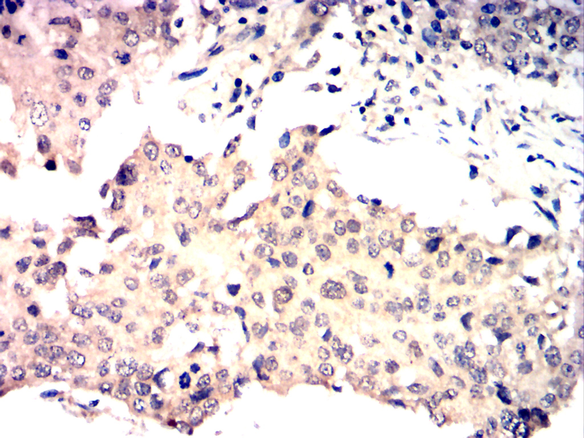
Immunohistochemical analysis of paraffin-embedded Human breast cancer tissues using CHK2 antibody with DAB staining.Pre-treat the sections with heat-mediated antigen retrieval using sodium citrate buffer (pH 6.0) (OM750020) for 2 minutes. Wash the sections with ddH₂O and PBS (OM750003). Block the tissue with 10% non-immune goat serum(OM760028) at room temperature for 30 minutes. Incubate the tissue with the primary antibody diluted at a ratio of 1:1500 at 4°C overnight. At room temperature, dilute the secondary antibody, Goat Anti-Mouse IgG (H&L) - HRP(OM644366), at a ratio of 1:200 and incubate for one hour. Use DAB(OM760029)as the chromogenic agent. Counterstain the tissue with hematoxylin, and mount the tissue sections with neutral gum.IHC
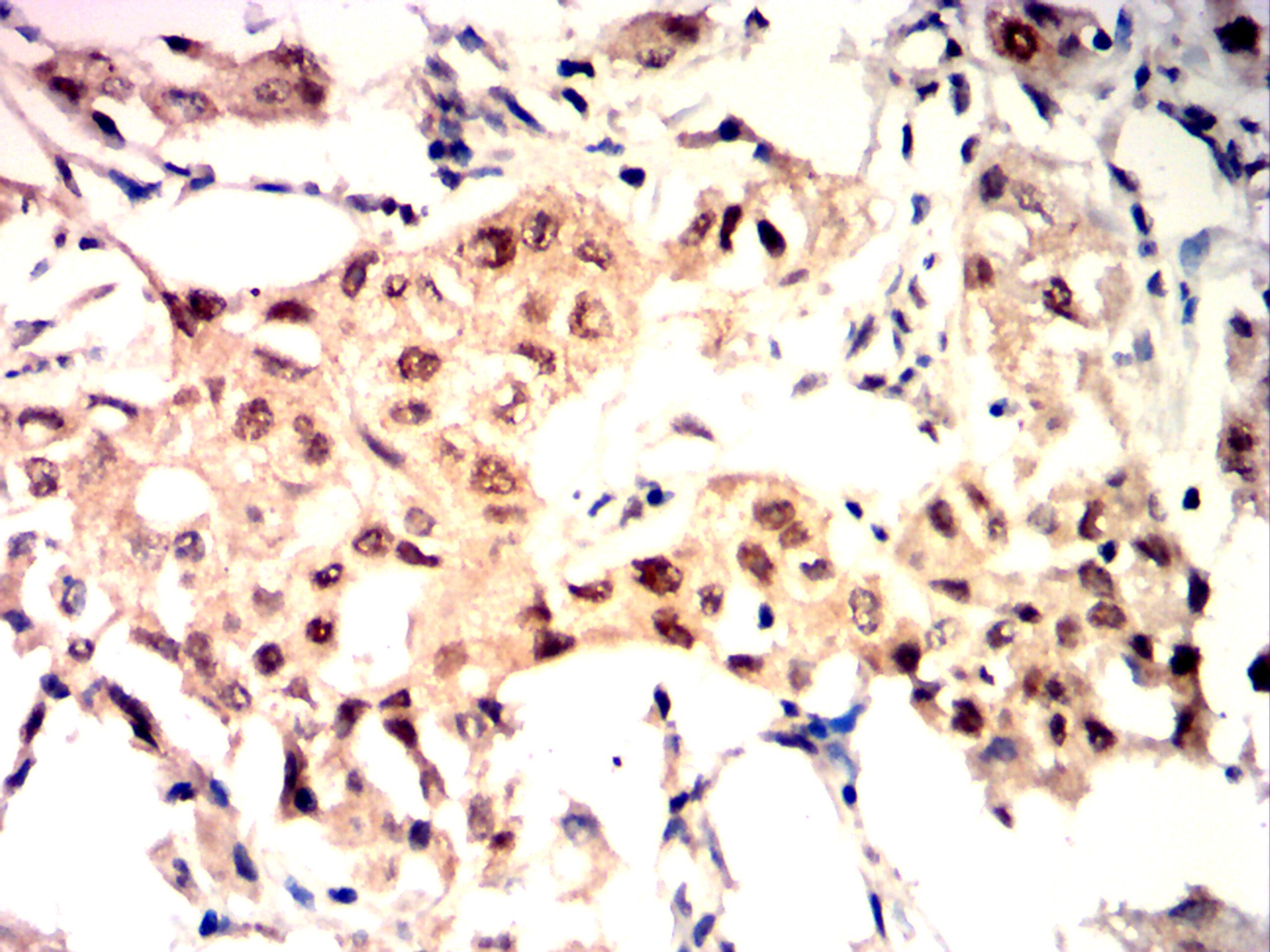
Immunohistochemical analysis of paraffin-embedded Human lung cancer tissues using CHK2 antibody with DAB staining.Pre-treat the sections with heat-mediated antigen retrieval using sodium citrate buffer (pH 6.0) (OM750020) for 2 minutes. Wash the sections with ddH₂O and PBS (OM750003). Block the tissue with 10% non-immune goat serum(OM760028) at room temperature for 30 minutes. Incubate the tissue with the primary antibody diluted at a ratio of 1:1500 at 4°C overnight. At room temperature, dilute the secondary antibody, Goat Anti-Mouse IgG (H&L) - HRP(OM644366), at a ratio of 1:200 and incubate for one hour. Use DAB(OM760029)as the chromogenic agent. Counterstain the tissue with hematoxylin, and mount the tissue sections with neutral gum.IHC
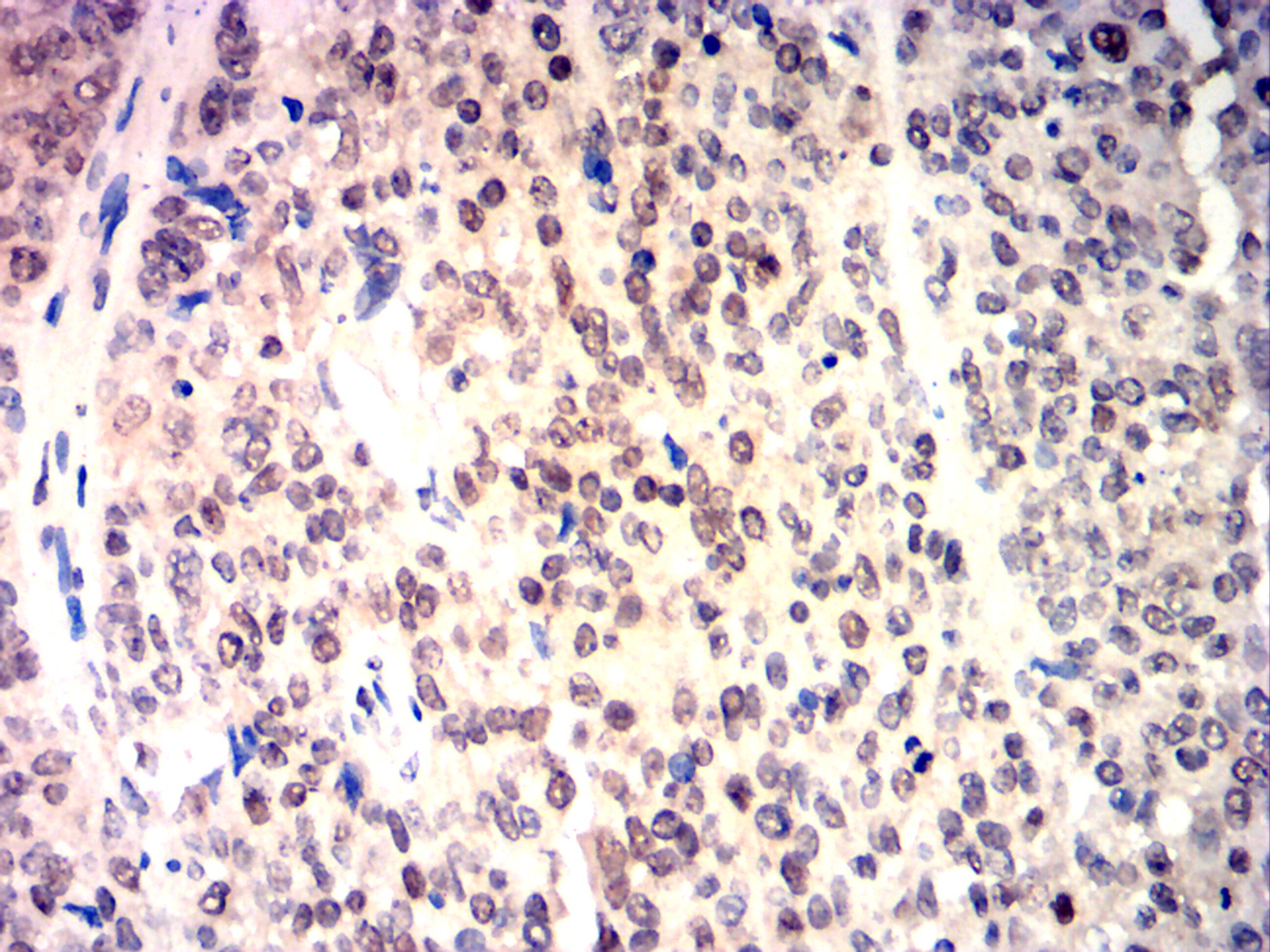
Immunohistochemical analysis of paraffin-embedded Human ovarian cancer tissues using CHK2 antibody with DAB staining.Pre-treat the sections with heat-mediated antigen retrieval using sodium citrate buffer (pH 6.0) (OM750020) for 2 minutes. Wash the sections with ddH₂O and PBS (OM750003). Block the tissue with 10% non-immune goat serum(OM760028) at room temperature for 30 minutes. Incubate the tissue with the primary antibody diluted at a ratio of 1:1500 at 4°C overnight. At room temperature, dilute the secondary antibody, Goat Anti-Mouse IgG (H&L) - HRP(OM644366), at a ratio of 1:200 and incubate for one hour. Use DAB(OM760029)as the chromogenic agent. Counterstain the tissue with hematoxylin, and mount the tissue sections with neutral gum.IHC
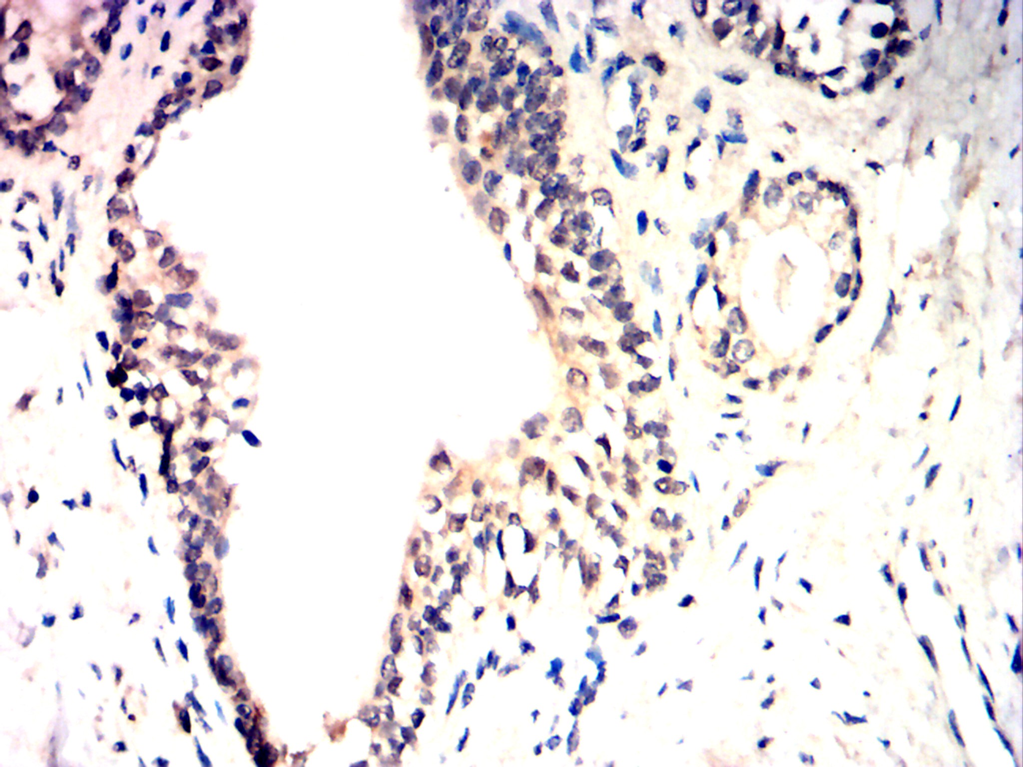
Immunohistochemical analysis of paraffin-embedded Human prostate tissues using CHK2 antibody with DAB staining.Pre-treat the sections with heat-mediated antigen retrieval using sodium citrate buffer (pH 6.0) (OM750020) for 2 minutes. Wash the sections with ddH₂O and PBS (OM750003). Block the tissue with 10% non-immune goat serum(OM760028) at room temperature for 30 minutes. Incubate the tissue with the primary antibody diluted at a ratio of 1:1500 at 4°C overnight. At room temperature, dilute the secondary antibody, Goat Anti-Mouse IgG (H&L) - HRP(OM644366), at a ratio of 1:200 and incubate for one hour. Use DAB(OM760029)as the chromogenic agent. Counterstain the tissue with hematoxylin, and mount the tissue sections with neutral gum.IHC
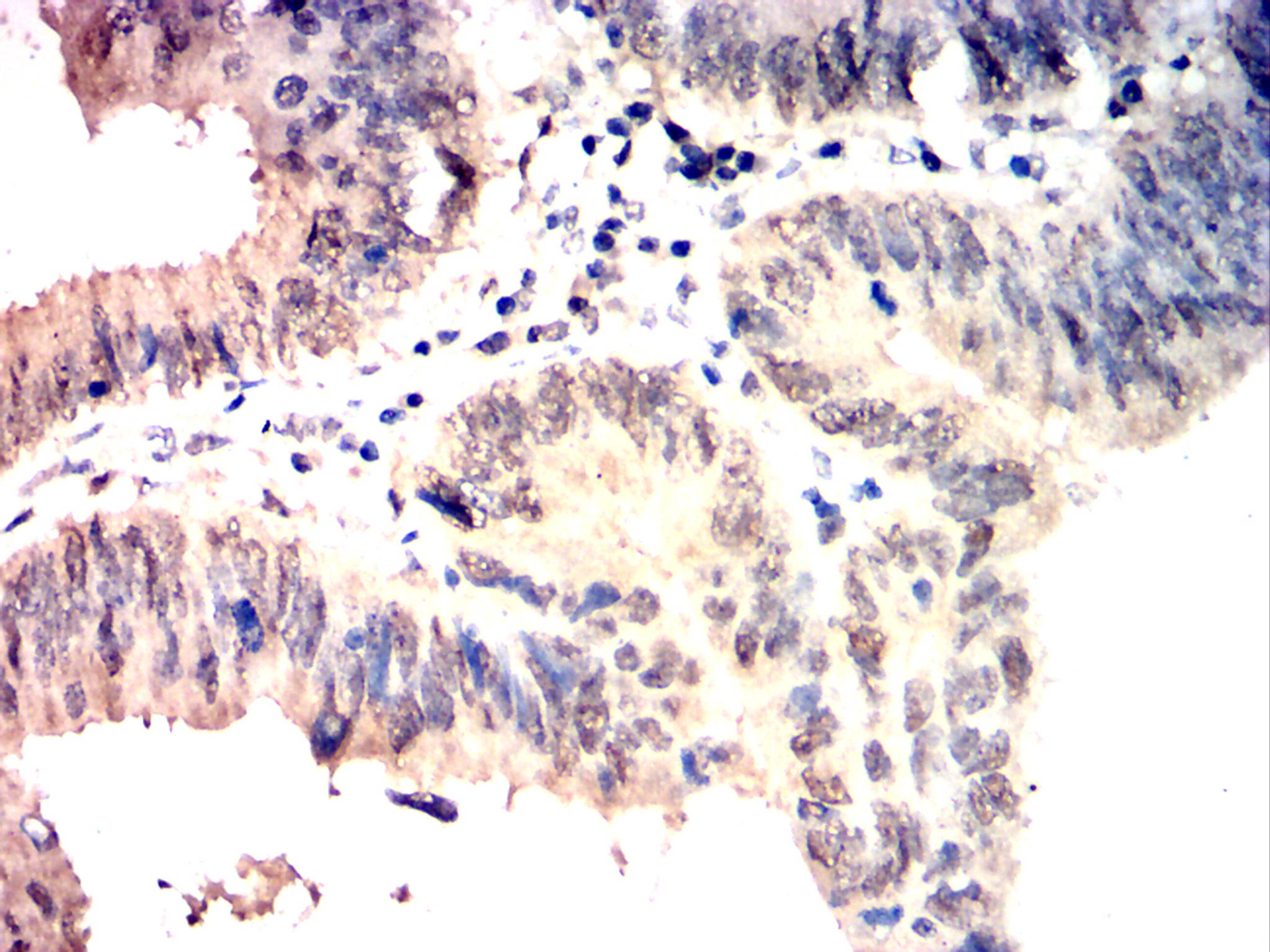
Immunohistochemical analysis of paraffin-embedded Human rectal cancer tissues using CHK2 antibody with DAB staining.Pre-treat the sections with heat-mediated antigen retrieval using sodium citrate buffer (pH 6.0) (OM750020) for 2 minutes. Wash the sections with ddH₂O and PBS (OM750003). Block the tissue with 10% non-immune goat serum(OM760028) at room temperature for 30 minutes. Incubate the tissue with the primary antibody diluted at a ratio of 1:1500 at 4°C overnight. At room temperature, dilute the secondary antibody, Goat Anti-Mouse IgG (H&L) - HRP(OM644366), at a ratio of 1:200 and incubate for one hour. Use DAB(OM760029)as the chromogenic agent. Counterstain the tissue with hematoxylin, and mount the tissue sections with neutral gum.ELISA
Coat the plate with immunizing antigen.Blue line: Antibody; Orange line: Isotype control antibody| Product Name | Anti-CHK2 antibody,pbs only |
|---|---|
| Antibody Type | Primary Antibodies |
| Immunogen | Recombinent Proteins |
| Clonality | Monoclonal |
|---|---|
| Isotype | IgG |
| Host Species | Mouse |
| Tested Applications | ELISAICC/IFIHCWB |
| WB:1:1000-1:2000 IHC:1:200-1:2000 ICC/IF:1:100-1:500 |
|
| Species Reactivity | HumanMouseRat |
| Concentration | 1mg/ml |
| Purification | Protein A |
| Gene Symbol | CHEK2 |
|---|---|
| Gene Synonyms | CDS1 CHK2 hCds1 HUCDS1 LFS2 PP1425 RAD53 TPDS4 |
| Gene Full Name | checkpoint kinase 2 |
| Gene Summary | In response to DNA damage and replication blocks, cell cycle progression is halted through the control of critical cell cycle regulators. The protein encoded by this gene is a cell cycle checkpoint regulator and putative tumor suppressor. It contains a forkhead-associated protein interaction domain essential for activation in response to DNA damage and is rapidly phosphorylated in response to replication blocks and DNA damage. When activated, the encoded protein is known to inhibit CDC25C phosphatase, preventing entry into mitosis, and has been shown to stabilize the tumor suppressor protein p53, leading to cell cycle arrest in G1. In addition, this protein interacts with and phosphorylates BRCA1, allowing BRCA1 to restore survival after DNA damage. Mutations in this gene have been linked with Li-Fraumeni syndrome, a highly penetrant familial cancer phenotype usually associated with inherited mutations in TP53. Also, mutations in this gene are thought to confer a predisposition to sarcomas, breast cancer, and brain tumors. This nuclear protein is a member of the CDS1 subfamily of serine/threonine protein kinases. Several transcript variants encoding different isoforms have been found for this gene. [provided by RefSeq, Apr 2012] |
| Alternative Names | Cds1 Cds1 homolog Checkpoint like protein CHK2 checkpoint-like protein CHK2 CHEK2 Chk2 (phospho T68) Chk2 (pThr68) CHK2 checkpoint homolog CHK2 checkpoint homolog (S. pombe) Chk2 phospho Chk2 phospho T68 Chk2-del2-12 Chk2-del2-3 Chk2-del4 Chk2-del7 Chk2-del9 Chk2-del9-12 Chk2-insX Chk2-insZ Chk2-iso1 Chk2-iso2 Chk2-sub3 contains FHA domain del9 EC 2.7.11.1 HuCds 1 kinase Chk2 OTTHUMP00000199044 OTTHUMP00000199045 OTTHUMP00000199064 OTTHUMP00000199115 OTTHUMP00000199116 phospho Chk2 (T68) phospho Thr68 Chk2 protein kinase Chk2 Rad53 homolog RP11-436C9.1 Serine/threonine protein kinase Chk2 |
| Molecular Weight(MW) | 61 kDa |
| Cellular Localization | Nucleus |
| SwissProt ID | O96017 |
|---|
WB
Western blot analysis using CHK2 antibody against HepG2 (1) cell lysate.12% SDS-PAGE gel.Sample loading: 20μg /lane. Transfer the proteins onto a PVDF membrane (OM790003), and block it with TBST (OM750016) plus skimmed milk powder(OM750084) for one hour. Dilute the primary antibody with the antibody diluent (OM750012) at a ratio of 1:1000, and incubate it overnight at 4°C. Wash the membrane three times with TBST (OM750016), 5 minutes each time. At room temperature, dilute the secondary antibody, Goat Anti-Mouse IgG (H&L) - HRP(OM644366), at a ratio of 1:20000 and incubate for one hour. Wash the membrane three times with TBST (OM750016) again, 5 minutes each time. Use ECL (OM625701) for luminescence.staining time: 60S.
IHC
Immunohistochemical analysis of paraffin-embedded Human prostate cancer tissues using CHK2 antibody with DAB staining.Pre-treat the sections with heat-mediated antigen retrieval using sodium citrate buffer (pH 6.0) (OM750020) for 2 minutes. Wash the sections with ddH₂O and PBS (OM750003). Block the tissue with 10% non-immune goat serum(OM760028) at room temperature for 30 minutes. Incubate the tissue with the primary antibody diluted at a ratio of 1:1500 at 4°C overnight. At room temperature, dilute the secondary antibody, Goat Anti-Mouse IgG (H&L) - HRP(OM644366), at a ratio of 1:200 and incubate for one hour. Use DAB(OM760029)as the chromogenic agent. Counterstain the tissue with hematoxylin, and mount the tissue sections with neutral gum.
ICC/IF
Immunofluorescence analysis of HepG2 cells using CHK2 antibody (green).Cells are fixed in 4% paraformaldehyde at room temperature for 20 minutes. Then, they are permeabilized with a PBS (OM750003) solution containing 0.1% Triton X-100(OM750021) at room temperature for 15 minutes. Subsequently, the cells are blocked with 10% non - immune goat serum(OM760028) at room temperature for 1 hour.The cells are incubated overnight at 4°C with the primary antibody diluted 1:100 in PBS. The secondary antibody, Omnimabs® 488 Goat Anti-Mouse IgG(H&L)(OM643485), is diluted at a ratio of 1:400 and incubated with the cells for 1 hour.Nuclear DNA is labeled with DAPI (Blue,OM643160). F-actin is stained with Omnimabs® 594-Phalloidin (Red,OM750007) diluted 1:100 for 30 minutes.
IHC
Immunohistochemical analysis of paraffin-embedded Human breast cancer tissues using CHK2 antibody with DAB staining.Pre-treat the sections with heat-mediated antigen retrieval using sodium citrate buffer (pH 6.0) (OM750020) for 2 minutes. Wash the sections with ddH₂O and PBS (OM750003). Block the tissue with 10% non-immune goat serum(OM760028) at room temperature for 30 minutes. Incubate the tissue with the primary antibody diluted at a ratio of 1:1500 at 4°C overnight. At room temperature, dilute the secondary antibody, Goat Anti-Mouse IgG (H&L) - HRP(OM644366), at a ratio of 1:200 and incubate for one hour. Use DAB(OM760029)as the chromogenic agent. Counterstain the tissue with hematoxylin, and mount the tissue sections with neutral gum.
IHC
Immunohistochemical analysis of paraffin-embedded Human lung cancer tissues using CHK2 antibody with DAB staining.Pre-treat the sections with heat-mediated antigen retrieval using sodium citrate buffer (pH 6.0) (OM750020) for 2 minutes. Wash the sections with ddH₂O and PBS (OM750003). Block the tissue with 10% non-immune goat serum(OM760028) at room temperature for 30 minutes. Incubate the tissue with the primary antibody diluted at a ratio of 1:1500 at 4°C overnight. At room temperature, dilute the secondary antibody, Goat Anti-Mouse IgG (H&L) - HRP(OM644366), at a ratio of 1:200 and incubate for one hour. Use DAB(OM760029)as the chromogenic agent. Counterstain the tissue with hematoxylin, and mount the tissue sections with neutral gum.
IHC
Immunohistochemical analysis of paraffin-embedded Human ovarian cancer tissues using CHK2 antibody with DAB staining.Pre-treat the sections with heat-mediated antigen retrieval using sodium citrate buffer (pH 6.0) (OM750020) for 2 minutes. Wash the sections with ddH₂O and PBS (OM750003). Block the tissue with 10% non-immune goat serum(OM760028) at room temperature for 30 minutes. Incubate the tissue with the primary antibody diluted at a ratio of 1:1500 at 4°C overnight. At room temperature, dilute the secondary antibody, Goat Anti-Mouse IgG (H&L) - HRP(OM644366), at a ratio of 1:200 and incubate for one hour. Use DAB(OM760029)as the chromogenic agent. Counterstain the tissue with hematoxylin, and mount the tissue sections with neutral gum.
IHC
Immunohistochemical analysis of paraffin-embedded Human prostate tissues using CHK2 antibody with DAB staining.Pre-treat the sections with heat-mediated antigen retrieval using sodium citrate buffer (pH 6.0) (OM750020) for 2 minutes. Wash the sections with ddH₂O and PBS (OM750003). Block the tissue with 10% non-immune goat serum(OM760028) at room temperature for 30 minutes. Incubate the tissue with the primary antibody diluted at a ratio of 1:1500 at 4°C overnight. At room temperature, dilute the secondary antibody, Goat Anti-Mouse IgG (H&L) - HRP(OM644366), at a ratio of 1:200 and incubate for one hour. Use DAB(OM760029)as the chromogenic agent. Counterstain the tissue with hematoxylin, and mount the tissue sections with neutral gum.
IHC
Immunohistochemical analysis of paraffin-embedded Human rectal cancer tissues using CHK2 antibody with DAB staining.Pre-treat the sections with heat-mediated antigen retrieval using sodium citrate buffer (pH 6.0) (OM750020) for 2 minutes. Wash the sections with ddH₂O and PBS (OM750003). Block the tissue with 10% non-immune goat serum(OM760028) at room temperature for 30 minutes. Incubate the tissue with the primary antibody diluted at a ratio of 1:1500 at 4°C overnight. At room temperature, dilute the secondary antibody, Goat Anti-Mouse IgG (H&L) - HRP(OM644366), at a ratio of 1:200 and incubate for one hour. Use DAB(OM760029)as the chromogenic agent. Counterstain the tissue with hematoxylin, and mount the tissue sections with neutral gum.
ELISA
Coat the plate with immunizing antigen.Blue line: Antibody; Orange line: Isotype control antibody| Application Notes | WB:1:1000-1:2000 IHC:1:200-1:2000 ICC/IF:1:100-1:500 |
|---|
| Form | Liquid |
|---|---|
| Storage Instructions | Shipped at 4°C. Store at +4°C short term (1-2 weeks). Store at -20°C long term. Avoid freeze / thaw cycle. |
| Storage Buffer | PBS ONLY |


