WB
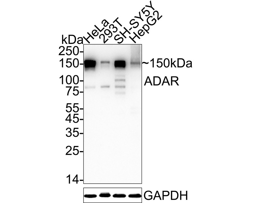
Western blot analysis of ADAR on different lysates with Rabbit anti-ADAR antibody at 1/1,000 dilution. Lane 1: HeLa cell lysate, Lane 2: 293T cell lysate, Lane 3: SH-SY5Y cell lysate, Lane 4: HepG2 cell lysate, Lysates/proteins at 20 µg/Lane. Exposure time: 1 minute; 4-20% SDS-PAGE gel. Proteins were transferred to a PVDF membrane and blocked with 5% NFDM/TBST for 1 hour at room temperature. The primary antibody at 1/1,000 dilution was used in 5% NFDM/TBST at 4℃ overnight. Goat Anti-Rabbit IgG - HRP Secondary Antibody at 1/50,000 dilution was used for 1 hour at room temperature.ICC/IF
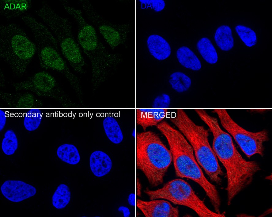
Immunocytochemistry analysis of HeLa cells labeling ADAR with Rabbit anti-ADAR antibody at 1/100 dilution. Cells were fixed in 4% paraformaldehyde for 20 minutes at room temperature, permeabilized with 0.1% Triton X-100 in PBS for 5 minutes at room temperature, then blocked with 1% BSA in 10% negative goat serum for 1 hour at room temperature. Cells were then incubated with Rabbit anti-ADAR antibody at 1/100 dilution in 1% BSA in PBST overnight at 4 ℃. Goat Anti-Rabbit IgG H&L (488) was used as the secondary antibody at 1/1,000 dilution. PBS instead of the primary antibody was used as the secondary antibody only control. Nuclear DNA was labelled in blue with DAPI. Beta tubulin (red) was stained at 1/100 dilution overnight at +4℃. Goat Anti-Mouse IgG H&L (594) was used as the secondary antibody at 1/1,000 dilution.IHC
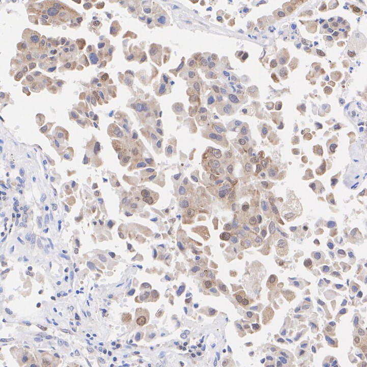
Immunohistochemical analysis of paraffin-embedded human lung cancer tissue with Rabbit anti-ADAR antibody at 1/1,000 dilution. The section was pre-treated using heat mediated antigen retrieval with sodium citrate buffer (pH 6.0) for 2 minutes. The tissues were blocked in 1% BSA for 20 minutes at room temperature, washed with ddH2O and PBS, and then probed with the primary antibody at 1/1,000 dilution for 1 hour at room temperature. The detection was performed using an HRP conjugated compact polymer system. DAB was used as the chromogen. Tissues were counterstained with hematoxylin and mounted with DPX.FC
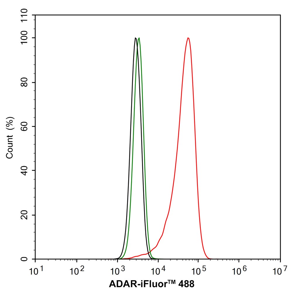
Flow cytometric analysis of HeLa cells labeling ADAR. Cells were fixed and permeabilized. Then stained with the primary antibody (1μg/mL) (red) compared with Rabbit IgG Isotype Control (green). After incubation of the primary antibody at +4℃ for an hour, the cells were stained with a iFluor™ 488 conjugate-Goat anti-Rabbit IgG Secondary antibody at 1/1,000 dilution for 30 minutes at +4℃. Unlabelled sample was used as a control (cells without incubation with primary antibody; black).| Product Name | ADAR Recombinant Rabbit Monoclonal Antibody |
|---|---|
| Antibody Type | Primary Antibodies |
| Product description | RNA-specific adenosine deaminase (ADAR1, DSH, IFI4, p136, DRADA, DSRAD, K88dsRBP) mediates RNA editing by destabilizing double stranded RNA through deamination of adenosine to inosine in structured or double-stranded RNAs. ADAR1 is expressed from an interferon-response promoter and has a Z-DNA/Z-RNA binding domain at its N-terminus. ADAR1 co-localizes with SUMO-1 in a subnucleolar region that is distinct from the fibrillar center, the dense fibrillar component and the granular component. Localization of nuclear ADAR1 is under the influence of a nucleolar localization signal (NoLS) in the middle of ADAR1 and the exporting activity of the nuclear exporter signal (NES) near the N terminus. ADAR1 upregulates nuclear factor 90 (NF90)-mediated gene expression by interacting with NF110, NF90 and NF45. ADAR1 binds short interfering RNA (siRNA), and gene silencing by siRNA is significantly more effective in mouse fibroblasts homozygous for an ADAR1 null mutation than in wild-type cells. ADAR1 may limit the efficacy of siRNA in mammalian cells. |
| Immunogen | Recombinant protein within Human ADAR aa 180-280 / 1,226. |
| Clonality | monoclonal |
|---|---|
| Isotype | IgG |
| Host Species | Recombinant rabbit |
| Tested Applications | FCICC/IFIHCWB |
| WB:1:1000 ICC/IF:1:100 IHC:1:200-1:1000 FC:1:1000 |
|
| Species Reactivity | Human |
| Concentration | 1mg/ml |
| Purification | Protein A |
| Gene Symbol | ADAR |
|---|---|
| Gene Synonyms | DSH AGS6 G1P1 IFI4 P136 ADAR1 DRADA DSRAD IFI-4 K88DSRBP |
| Gene Full Name | adenosine deaminase RNA specific |
| Molecular Weight(MW) | 136kDa(Observed band size: 150kDa) |
| Cellular Localization | Cytoplasm. Nucleus. |
| SwissProt ID | P55265 |
|---|
WB
Western blot analysis of ADAR on different lysates with Rabbit anti-ADAR antibody at 1/1,000 dilution. Lane 1: HeLa cell lysate, Lane 2: 293T cell lysate, Lane 3: SH-SY5Y cell lysate, Lane 4: HepG2 cell lysate, Lysates/proteins at 20 µg/Lane. Exposure time: 1 minute; 4-20% SDS-PAGE gel. Proteins were transferred to a PVDF membrane and blocked with 5% NFDM/TBST for 1 hour at room temperature. The primary antibody at 1/1,000 dilution was used in 5% NFDM/TBST at 4℃ overnight. Goat Anti-Rabbit IgG - HRP Secondary Antibody at 1/50,000 dilution was used for 1 hour at room temperature.
ICC/IF
Immunocytochemistry analysis of HeLa cells labeling ADAR with Rabbit anti-ADAR antibody at 1/100 dilution. Cells were fixed in 4% paraformaldehyde for 20 minutes at room temperature, permeabilized with 0.1% Triton X-100 in PBS for 5 minutes at room temperature, then blocked with 1% BSA in 10% negative goat serum for 1 hour at room temperature. Cells were then incubated with Rabbit anti-ADAR antibody at 1/100 dilution in 1% BSA in PBST overnight at 4 ℃. Goat Anti-Rabbit IgG H&L (488) was used as the secondary antibody at 1/1,000 dilution. PBS instead of the primary antibody was used as the secondary antibody only control. Nuclear DNA was labelled in blue with DAPI. Beta tubulin (red) was stained at 1/100 dilution overnight at +4℃. Goat Anti-Mouse IgG H&L (594) was used as the secondary antibody at 1/1,000 dilution.
IHC
Immunohistochemical analysis of paraffin-embedded human lung cancer tissue with Rabbit anti-ADAR antibody at 1/1,000 dilution. The section was pre-treated using heat mediated antigen retrieval with sodium citrate buffer (pH 6.0) for 2 minutes. The tissues were blocked in 1% BSA for 20 minutes at room temperature, washed with ddH2O and PBS, and then probed with the primary antibody at 1/1,000 dilution for 1 hour at room temperature. The detection was performed using an HRP conjugated compact polymer system. DAB was used as the chromogen. Tissues were counterstained with hematoxylin and mounted with DPX.
FC
Flow cytometric analysis of HeLa cells labeling ADAR. Cells were fixed and permeabilized. Then stained with the primary antibody (1μg/mL) (red) compared with Rabbit IgG Isotype Control (green). After incubation of the primary antibody at +4℃ for an hour, the cells were stained with a iFluor™ 488 conjugate-Goat anti-Rabbit IgG Secondary antibody at 1/1,000 dilution for 30 minutes at +4℃. Unlabelled sample was used as a control (cells without incubation with primary antibody; black).| Positive Control | Hela, human lung cancer tissue, human colon cancer tissue, human kidney tissue, human pancreas tissue, SiHa. |
|---|---|
| Application Notes | WB:1:1000 ICC/IF:1:100 IHC:1:200-1:1000 FC:1:1000 |
| Form | Liquid |
|---|---|
| Storage Instructions | Store at +4℃ after thawing. Aliquot store at -20℃ or -80℃. Avoid repeated freeze / thaw cycles. |
| Storage Buffer | 1*TBS (pH7.4), 1%BSA, 40%Glycerol. Preservative: 0.05% Sodium Azide. |


