WB
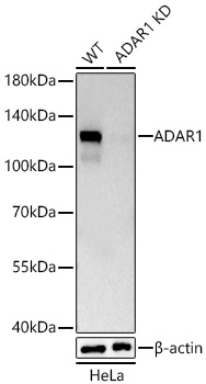
Western blot analysis of lysates from wild type (WT) and ADAR1 knockdown (KD) HeLa cells using [KDValidated] ADAR1 Rabbit mAb at 1:5000 dilution incubated at room temperature for 1.5 hours.Secondary antibody: HRP-conjugated Goat anti-Rabbit IgG (H+L) at 1:10000 dilution.Lysates/proteins: 25 μg per lane.Blocking buffer: 3% nonfat dry milk in TBST.Detection: ECL Basic Kit.Exposure time: 10s.ICC/IF
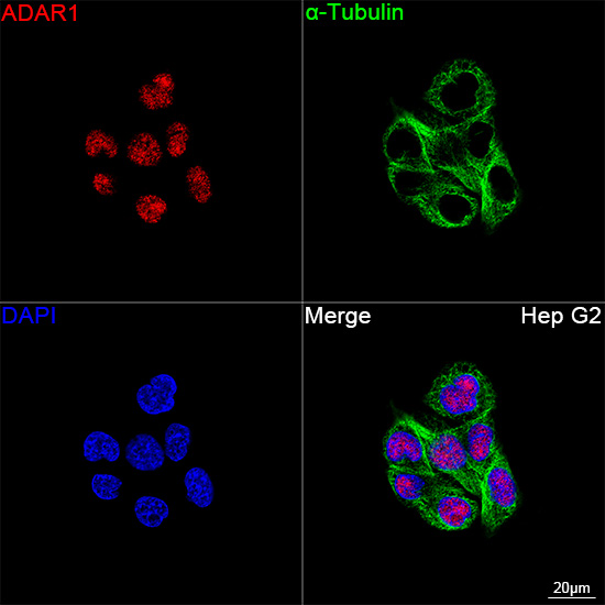
Confocal imaging of Hep G2 cells using [KDValidated] ADAR1 Rabbit mAb (dilution 1:200) followed by a furtherincubation with Cy3 Goat Anti-Rabbit IgG(H+L) (dilution 1:500) (Red). Thecells were counterstained with α-TubulinMouse mAb (dilution 1:400) followedby incubation with ABflo® 488-conjugatedGoat Anti-Mouse IgG (H+L) Ab (dilution 1:500) (Green). DAPI was used fornuclear staining (Blue). Objective: 100x.ICC/IF
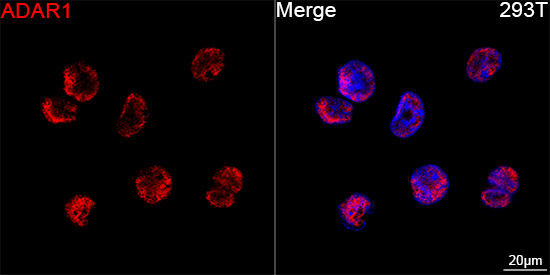
Confocal imaging of 293T cells using [KDValidated] ADAR1 Rabbit mAb (dilution 1:200) followed by a furtherincubation with Cy3 Goat Anti-Rabbit IgG(H+L) (dilution 1:500) (Red). DAPIwas used for nuclear staining (Blue).Objective: 100x.IHC
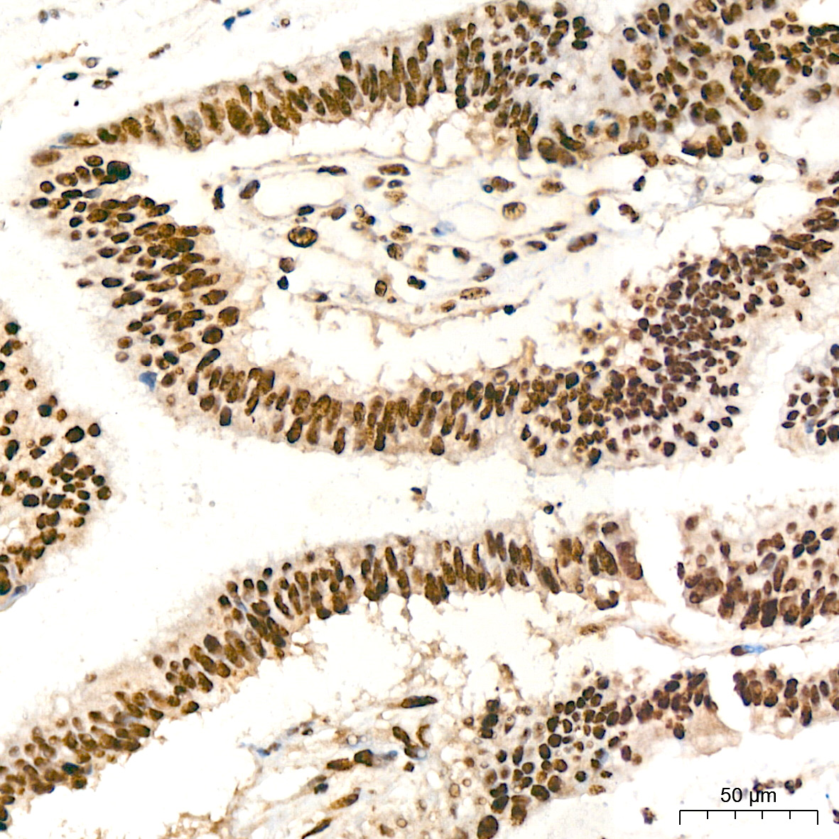
Immunohistochemistry analysis of paraffinembedded Human cervix cancer tissue using[KD Validated] ADAR1 Rabbit mAb at a dilution of 1:200 (40x lens). Highpressure antigen retrieval performed with0.01M Citrate Buffer (pH 6.0) prior to IHCstaining.| Product Name | [KD Validated] ADAR1 Rabbit mAb |
|---|---|
| Antibody Type | Primary Antibodies |
| Immunogen | Recombinant protein (or fragment).This information is considered to be commercially sensitive. |
| Clonality | Monoclonal |
|---|---|
| Isotype | IgG |
| Host Species | Rabbit |
| Tested Applications | ICC/IFIHCWB |
| WB:1:5000-1:30000 ICC/IF:1:100-1:400 IHC:1:200-1:2000 |
|
| Species Reactivity | Human |
| Concentration | 1mg/ml |
| Purification | Affinity purified |
| Gene Symbol | ADAR |
|---|---|
| Gene Synonyms | DSH AGS6 G1P1 IFI4 P136 ADAR1 DRADA DSRAD IFI-4 K88DSRBP |
| Gene Full Name | adenosine deaminase RNA specific |
| Gene Summary | This gene encodes the enzyme responsible for RNA editing by site-specific deamination of adenosines. This enzyme destabilizes double-stranded RNA through conversion of adenosine to inosine. Mutations in this gene have been associated with dyschromatosis symmetrica hereditaria. Alternative splicing results in multiple transcript variants. [provided by RefSeq, Jul 2010] |
| Alternative Names | DSH AGS6 G1P1 IFI4 P136 ADAR1 DRADA DSRAD IFI-4 K88DSRBP |
| Molecular Weight(MW) | 136kDa(Observed band size:110kDa) |
| Cellular Localization | Cytoplasm, Nucleus, Nucleus, nucleolus. |
WB
Western blot analysis of lysates from wild type (WT) and ADAR1 knockdown (KD) HeLa cells using [KDValidated] ADAR1 Rabbit mAb at 1:5000 dilution incubated at room temperature for 1.5 hours.Secondary antibody: HRP-conjugated Goat anti-Rabbit IgG (H+L) at 1:10000 dilution.Lysates/proteins: 25 μg per lane.Blocking buffer: 3% nonfat dry milk in TBST.Detection: ECL Basic Kit.Exposure time: 10s.
ICC/IF
Confocal imaging of Hep G2 cells using [KDValidated] ADAR1 Rabbit mAb (dilution 1:200) followed by a furtherincubation with Cy3 Goat Anti-Rabbit IgG(H+L) (dilution 1:500) (Red). Thecells were counterstained with α-TubulinMouse mAb (dilution 1:400) followedby incubation with ABflo® 488-conjugatedGoat Anti-Mouse IgG (H+L) Ab (dilution 1:500) (Green). DAPI was used fornuclear staining (Blue). Objective: 100x.
ICC/IF
Confocal imaging of 293T cells using [KDValidated] ADAR1 Rabbit mAb (dilution 1:200) followed by a furtherincubation with Cy3 Goat Anti-Rabbit IgG(H+L) (dilution 1:500) (Red). DAPIwas used for nuclear staining (Blue).Objective: 100x.
IHC
Immunohistochemistry analysis of paraffinembedded Human cervix cancer tissue using[KD Validated] ADAR1 Rabbit mAb at a dilution of 1:200 (40x lens). Highpressure antigen retrieval performed with0.01M Citrate Buffer (pH 6.0) prior to IHCstaining.| Application Notes | WB:1:5000-1:30000 ICC/IF:1:100-1:400 IHC:1:200-1:2000 |
|---|
| Form | Liquid |
|---|---|
| Storage Instructions | Store at -20℃. Avoid freeze / thaw cycles. |
| Storage Buffer | PBS containing 50% glycerol and 0.05% BSA, preserved with proclin300 or sodium azide (as specified on the Certificate of Analysis), pH 7.3. |


