WB
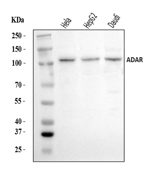
Western blot analysis of ADAR using anti-ADAR antibody. The sample well of each lane was loaded with 30 ug of sample under reducing conditions. Lane 1: HELA whole cell lysates, Lane 2: HEPG2 whole cell lysates, Lane 3: Daudi whole cell lysates. After electrophoresis, proteins were transferred to a membrane. Then the membrane was incubated with rabbit anti-ADAR antigen affinity purified polyclonal antibody at a dilution of 1:1000 and probed with a goat anti-rabbit IgG-HRP secondary antibody. The signal is developed using ECL Plus Western Blotting Substrate.IHC
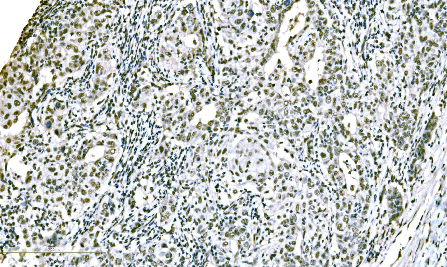
ADAR was detected in a paraffin-embedded section of human the renal pelvis is squamous metaplasia tissue. Biotinylated goat anti-rabbit IgG was used as secondary antibody. The tissue section was incubated with rabbit anti-ADAR Antibody at a dilution of 1:200 and developed using Strepavidin-Biotin-Complex (SABC) with DAB as the chromogen.IHC
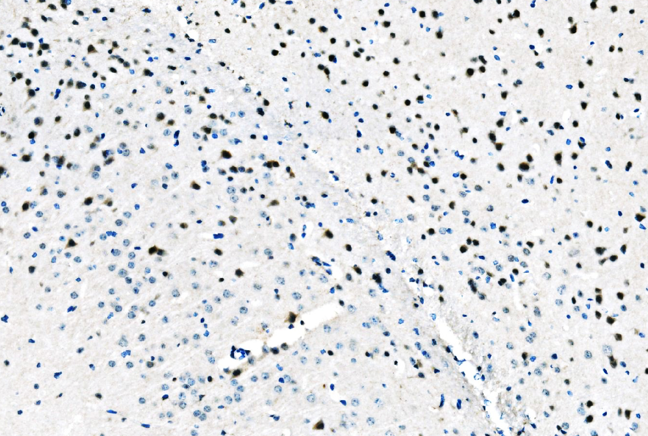
ADAR was detected in a paraffin-embedded section of mouse brain tissue. Biotinylated goat anti-rabbit IgG was used as secondary antibody. The tissue section was incubated with rabbit anti-ADAR Antibody at a dilution of 1:200 and developed using Strepavidin-Biotin-Complex (SABC) with DAB as the chromogen.IHC
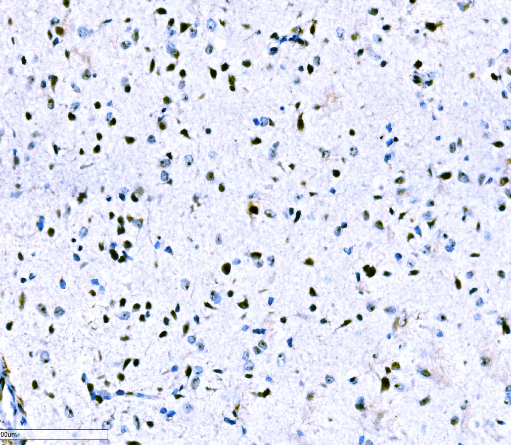
ADAR was detected in a paraffin-embedded section of rat brain tissue. Biotinylated goat anti-rabbit IgG was used as secondary antibody. The tissue section was incubated with rabbit anti-ADAR Antibody at a dilution of 1:200 and developed using Strepavidin-Biotin-Complex (SABC) with DAB as the chromogen.ICC/IF
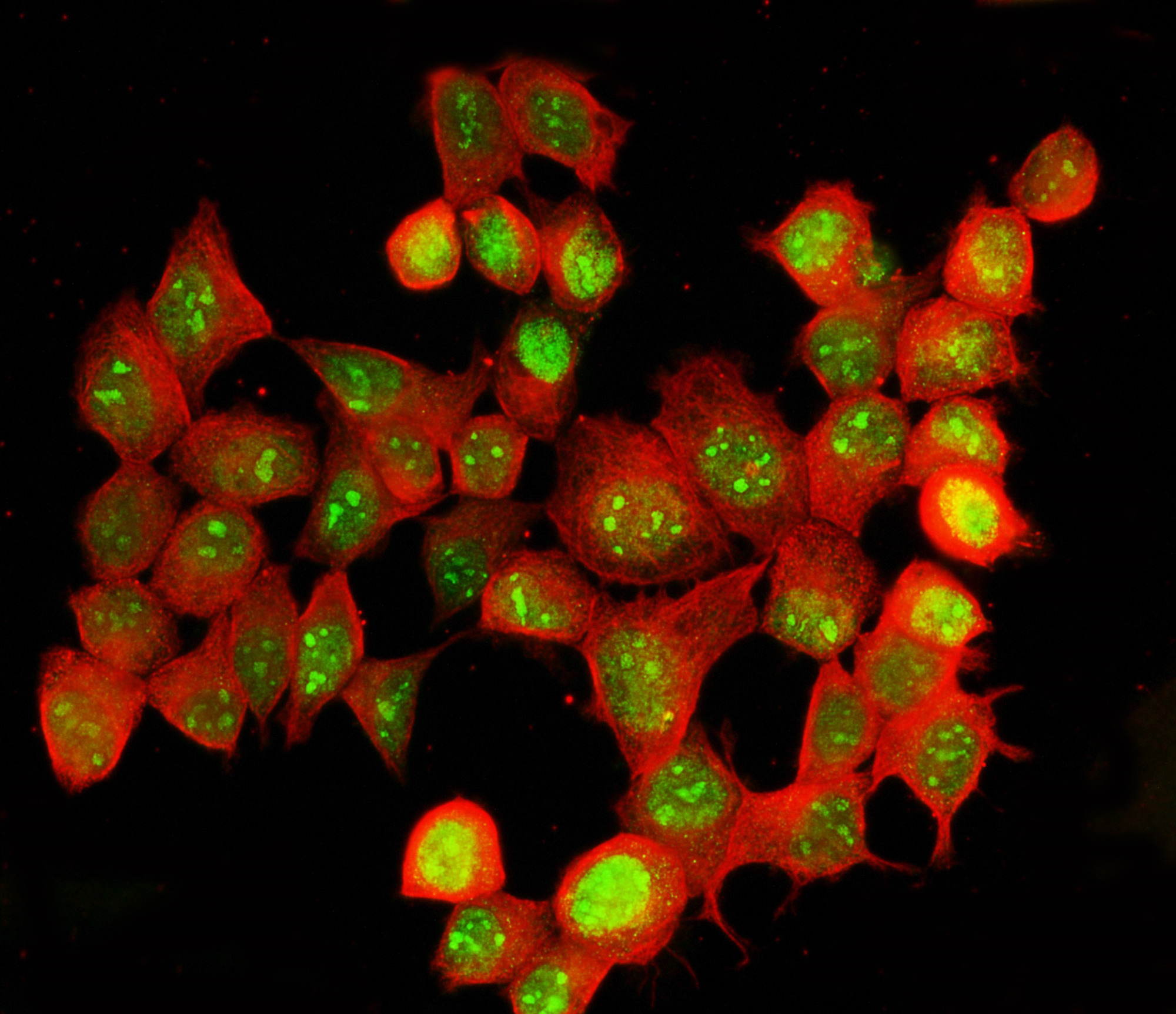
ADAR was detected in an immunocytochemical section of MCF-7 cells. The section was incubated with rabbit anti-ADAR Antibody at a dilution of 1:100. Fluoro488-conjugated Anti-rabbit IgG Secondary Antibody (green) and Fluoro594-conjugated Anti-mouse IgG Secondary Antibody (red) were used as secondary antibody.| Product Name | Rabbit polyclonal antibody to ADAR |
|---|---|
| Antibody Type | Primary Antibodies |
| Immunogen | E.coli-derived human ADAR1/ADAR recombinant protein (Position: M1-E1152). |
| Clonality | Polyclonal |
|---|---|
| Isotype | IgG |
| Host Species | Rabbit |
| Tested Applications | ICC/IFIHCWB |
| WB:1:500-1:2000 IHC:1:50-1:400 ICC/IF:1:50-1:400 |
|
| Species Reactivity | HumanMouseRat |
| Concentration | 0.5mg/ml |
| Purification | Affinity purified |
| Gene Symbol | ADAR |
|---|---|
| Gene Synonyms | DSH AGS6 G1P1 IFI4 P136 ADAR1 DRADA DSRAD IFI-4 K88DSRBP |
| Gene Full Name | adenosine deaminase RNA specific |
| Gene Summary | This gene encodes the enzyme responsible for RNA editing by site-specific deamination of adenosines. This enzyme destabilizes double-stranded RNA through conversion of adenosine to inosine. Mutations in this gene have been associated with dyschromatosis symmetrica hereditaria. Alternative splicing results in multiple transcript variants. [provided by RefSeq, Jul 2010] |
| Alternative Names | ADAR ADAR1 adenosine deaminase RNA DRADA DSH DSRAD G1P1 IFI 4 IFI4 K88dsRBP p136 |
| Molecular Weight(MW) | 136 kDa(Observed MW 110 kDa/150 kDa) |
| Cellular Localization | Cytoplasm,Nucleus. |
WB
Western blot analysis of ADAR using anti-ADAR antibody. The sample well of each lane was loaded with 30 ug of sample under reducing conditions. Lane 1: HELA whole cell lysates, Lane 2: HEPG2 whole cell lysates, Lane 3: Daudi whole cell lysates. After electrophoresis, proteins were transferred to a membrane. Then the membrane was incubated with rabbit anti-ADAR antigen affinity purified polyclonal antibody at a dilution of 1:1000 and probed with a goat anti-rabbit IgG-HRP secondary antibody. The signal is developed using ECL Plus Western Blotting Substrate.
IHC
ADAR was detected in a paraffin-embedded section of human the renal pelvis is squamous metaplasia tissue. Biotinylated goat anti-rabbit IgG was used as secondary antibody. The tissue section was incubated with rabbit anti-ADAR Antibody at a dilution of 1:200 and developed using Strepavidin-Biotin-Complex (SABC) with DAB as the chromogen.
IHC
ADAR was detected in a paraffin-embedded section of mouse brain tissue. Biotinylated goat anti-rabbit IgG was used as secondary antibody. The tissue section was incubated with rabbit anti-ADAR Antibody at a dilution of 1:200 and developed using Strepavidin-Biotin-Complex (SABC) with DAB as the chromogen.
IHC
ADAR was detected in a paraffin-embedded section of rat brain tissue. Biotinylated goat anti-rabbit IgG was used as secondary antibody. The tissue section was incubated with rabbit anti-ADAR Antibody at a dilution of 1:200 and developed using Strepavidin-Biotin-Complex (SABC) with DAB as the chromogen.
ICC/IF
ADAR was detected in an immunocytochemical section of MCF-7 cells. The section was incubated with rabbit anti-ADAR Antibody at a dilution of 1:100. Fluoro488-conjugated Anti-rabbit IgG Secondary Antibody (green) and Fluoro594-conjugated Anti-mouse IgG Secondary Antibody (red) were used as secondary antibody.| Application Notes | WB:1:500-1:2000 IHC:1:50-1:400 ICC/IF:1:50-1:400 |
|---|
| Form | Liquid |
|---|---|
| Storage Instructions | 12 months from date of receipt, -20℃ as supplied. 6 months 2 to 8℃ after reconstitution. Avoid repeated freezing and thawing. |
| Storage Buffer | 500ug/ml antibody with PBS, 0.02% NaN3, 1 mg/ml BSA and 50% glycerol. |


