WB
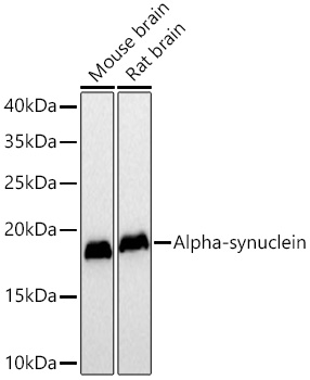
Western blot analysis of various lysates using Alpha-synuclein Mouse mAb at 1:5000 dilution incubated overnight at 4℃. Secondary antibody: HRP-conjugated Goat anti-Mouse IgG (H+L) at 1:10000 dilution. Lysates/proteins: 25 μg per lane. Blocking buffer: 3% nonfat dry milk in TBST. Detection: ECL Basic Kit. Exposure time: 90s.IHC
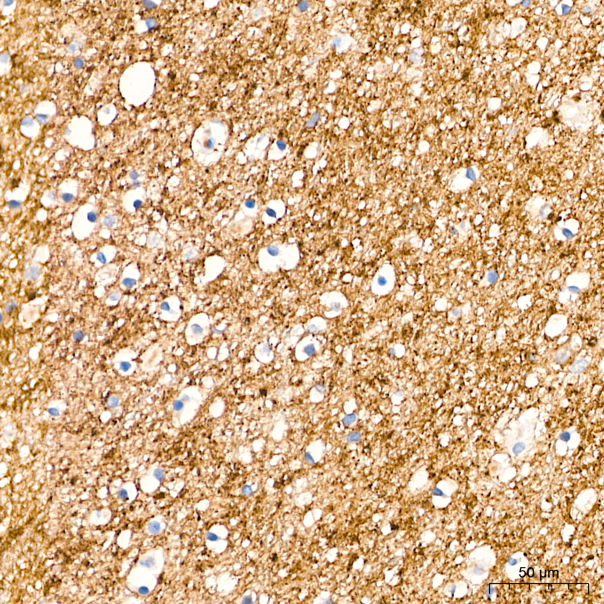
Immunohistochemistry analysis of paraffin embedded Human brain tissue using Alpha synuclein Mouse mAb at a dilution of 1:2000 (40x lens). High pressure antigen retrieval performed with 0.01M Citrate Buffer (pH 6.0) prior to IHC staining.IHC
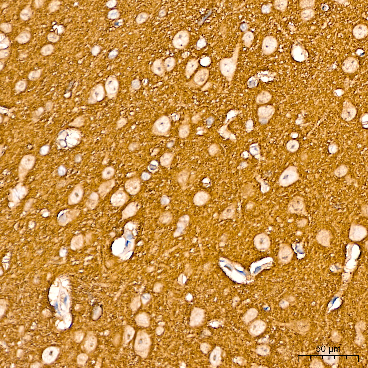
Immunohistochemistry analysis of paraffin embedded Mouse brain tissue using Alpha synuclein Mouse mAb at a dilution of 1:2000 (40x lens). High pressure antigen retrieval performed with 0.01M Citrate Buffer (pH 6.0) prior to IHC staining.IHC
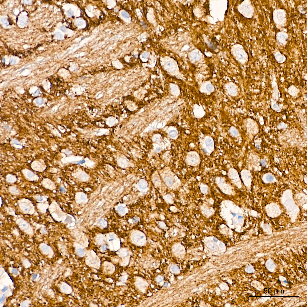
Immunohistochemistry analysis of paraffin embedded Rat brain tissue using Alpha synuclein Mouse mAb at a dilution of 1:2000 (40x lens). High pressure antigen retrieval performed with 0.01M Citrate Buffer (pH 6.0) prior to IHC staining.ICC/IF
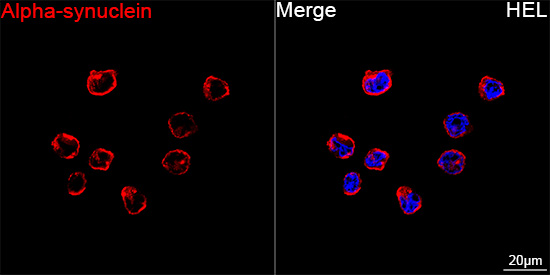
Confocal imaging of HEL cells using Alpha synuclein Mouse mAb (dilution 1:200) followed by a further incubation with Cy3 Goat Anti-Mouse IgG (H+L) (dilution 1:500) (Red). DAPI was used for nuclear staining (Blue). Objective: 100x.IF-P
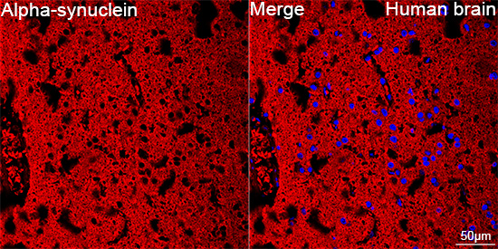
Confocal imaging of paraffin-embedded Human brain tissue using Alpha-synuclein Mouse mAb (dilution 1:200) followed by a further incubation with Cy3 Goat Anti-Mouse IgG (H+L) (dilution 1:500) (Red). DAPI was used for nuclear staining (Blue). Microwave antigen retrieval performed with 0.01M Citrate Buffer (pH 6.0) prior to IF staining. Objective: 40x.IF-P
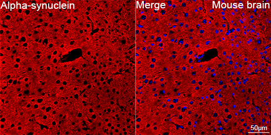
Confocal imaging of paraffin-embedded Mouse brain tissue using Alpha-synuclein Mouse mAb (dilution 1:200) followed by a further incubation with Cy3 conjugated Goat anti-Mouse IgG (H+L) (dilution 1:500) (Red). DAPI was used for nuclear staining (Blue). Microwave antigen retrieval performed with 0.01M Citrate Buffer (pH 6.0) prior to IF staining. Objective: 40x.IF-P
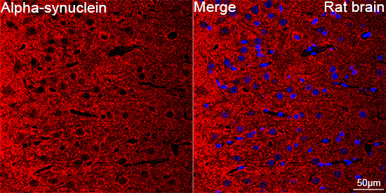
Confocal imaging of paraffin-embedded Rat brain tissue using Alpha-synuclein Mouse mAb (dilution 1:200) followed by a further incubation with Cy3 Goat Anti-Mouse IgG (H+L) (dilution 1:500) (Red). DAPI was used for nuclear staining (Blue). Microwave antigen retrieval performed with 0.01M Citrate Buffer (pH 6.0) prior to IF staining. Objective: 40x.| Product Name | Alpha-synuclein Mouse mAb |
|---|---|
| Antibody Type | Primary Antibodies |
| Immunogen | Recombinant fusion protein containing a sequence corresponding to amino acids 1-140 of human Alpha-synuclein(NP_000336.1). |
| Clonality | Monoclonal |
|---|---|
| Isotype | IgG |
| Host Species | Mouse |
| Tested Applications | ICC/IFIF-PIHCWB |
| WB:1:2500-1:10000 IHC:1:1000-1:4000 ICC/IF:1:200-1:800 IF-P:1:200-1:800 |
|
| Species Reactivity | HumanMouseRat |
| Concentration | 1mg/ml |
| Purification | Affinity purified |
| Gene Symbol | SNCA |
|---|---|
| Gene Synonyms | PD1 NACP PARK1 PARK4 |
| Gene Full Name | synuclein alpha |
| Gene Summary | Alpha-synuclein is a member of the synuclein family, which also includes beta- and gamma-synuclein. Synucleins are abundantly expressed in the brain and alpha- and beta-synuclein inhibit phospholipase D2 selectively. SNCA may serve to integrate presynaptic signaling and membrane trafficking. Defects in SNCA have been implicated in the pathogenesis of Parkinson disease. SNCA peptides are a major component of amyloid plaques in the brains of patients with Alzheimer's disease. Alternatively spliced transcripts encoding different isoforms have been identified for this gene. [provided by RefSeq, Feb 2016] |
| Alternative Names | PD1 NACP PARK1 PARK4 |
| Molecular Weight(MW) | 14kDa(Observed MW 18kDa) |
| Cellular Localization | Cell junction, Cytoplasm, Membrane, Nucleus, Secreted, cytosol, synapse, Cell projection, axon. |
WB
Western blot analysis of various lysates using Alpha-synuclein Mouse mAb at 1:5000 dilution incubated overnight at 4℃. Secondary antibody: HRP-conjugated Goat anti-Mouse IgG (H+L) at 1:10000 dilution. Lysates/proteins: 25 μg per lane. Blocking buffer: 3% nonfat dry milk in TBST. Detection: ECL Basic Kit. Exposure time: 90s.
IHC
Immunohistochemistry analysis of paraffin embedded Human brain tissue using Alpha synuclein Mouse mAb at a dilution of 1:2000 (40x lens). High pressure antigen retrieval performed with 0.01M Citrate Buffer (pH 6.0) prior to IHC staining.
IHC
Immunohistochemistry analysis of paraffin embedded Mouse brain tissue using Alpha synuclein Mouse mAb at a dilution of 1:2000 (40x lens). High pressure antigen retrieval performed with 0.01M Citrate Buffer (pH 6.0) prior to IHC staining.
IHC
Immunohistochemistry analysis of paraffin embedded Rat brain tissue using Alpha synuclein Mouse mAb at a dilution of 1:2000 (40x lens). High pressure antigen retrieval performed with 0.01M Citrate Buffer (pH 6.0) prior to IHC staining.
ICC/IF
Confocal imaging of HEL cells using Alpha synuclein Mouse mAb (dilution 1:200) followed by a further incubation with Cy3 Goat Anti-Mouse IgG (H+L) (dilution 1:500) (Red). DAPI was used for nuclear staining (Blue). Objective: 100x.
IF-P
Confocal imaging of paraffin-embedded Human brain tissue using Alpha-synuclein Mouse mAb (dilution 1:200) followed by a further incubation with Cy3 Goat Anti-Mouse IgG (H+L) (dilution 1:500) (Red). DAPI was used for nuclear staining (Blue). Microwave antigen retrieval performed with 0.01M Citrate Buffer (pH 6.0) prior to IF staining. Objective: 40x.
IF-P
Confocal imaging of paraffin-embedded Mouse brain tissue using Alpha-synuclein Mouse mAb (dilution 1:200) followed by a further incubation with Cy3 conjugated Goat anti-Mouse IgG (H+L) (dilution 1:500) (Red). DAPI was used for nuclear staining (Blue). Microwave antigen retrieval performed with 0.01M Citrate Buffer (pH 6.0) prior to IF staining. Objective: 40x.
IF-P
Confocal imaging of paraffin-embedded Rat brain tissue using Alpha-synuclein Mouse mAb (dilution 1:200) followed by a further incubation with Cy3 Goat Anti-Mouse IgG (H+L) (dilution 1:500) (Red). DAPI was used for nuclear staining (Blue). Microwave antigen retrieval performed with 0.01M Citrate Buffer (pH 6.0) prior to IF staining. Objective: 40x.| Application Notes | WB:1:2500-1:10000 IHC:1:1000-1:4000 ICC/IF:1:200-1:800 IF-P:1:200-1:800 |
|---|
| Form | Liquid |
|---|---|
| Storage Instructions | Store at -20℃. Avoid freeze / thaw cycles. |
| Storage Buffer | PBS with 0.02% sodium azide, 0.05% BSA, 50% glycerol, pH7.3. |


