WB
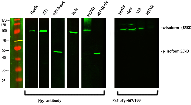
Western Blot analysis of various cells using primary antibody diluted at 1:1000(4°C overnight). Secondary antibody:Goat Anti-rabbit IgG IRDye 800( diluted at 1:5000, 25°C, 1 hour)WB
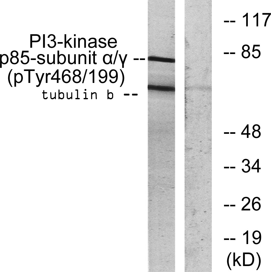
Western blot analysis of lysates from COS7 cells treated with H2O2 100uM 30', using PI3-kinase p85-alpha/gamma (Phospho-Tyr467/199) Antibody. The lane on the right is blocked with the phospho peptide.IHC
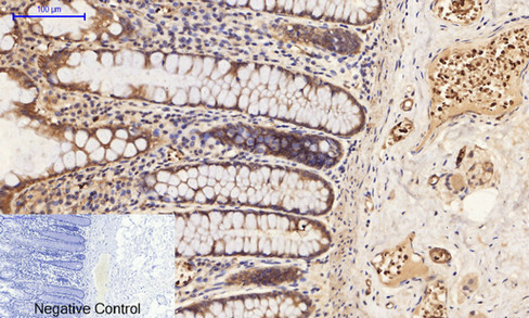
Immunohistochemical analysis of paraffin-embedded Human-colon tissue. 1,PI 3-kinase p85/p55 (phospho Tyr467/199) Polyclonal Antibody was diluted at 1:200(4°C,overnight). 2, Sodium citrate pH 6.0 was used for antibody retrieval(>98°C,20min). 3,Secondary antibody was diluted at 1:200(room tempeRature, 30min). Negative control was used by secondary antibody only.IHC
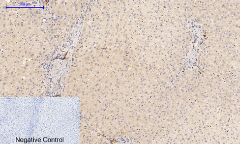
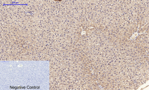
Immunohistochemical analysis of paraffin-embedded Human-liver tissue. 1,PI 3-kinase p85/p55 (phospho Tyr467/199) Polyclonal Antibody was diluted at 1:200(4°C,overnight). 2, Sodium citrate pH 6.0 was used for antibody retrieval(>98°C,20min). 3,Secondary antibody was diluted at 1:200(room tempeRature, 30min). Negative control was used by secondary antibody only.IHC
Immunohistochemical analysis of paraffin-embedded Rat-liver tissue. 1,PI 3-kinase p85/p55 (phospho Tyr467/199) Polyclonal Antibody was diluted at 1:200(4°C,overnight). 2, Sodium citrate pH 6.0 was used for antibody retrieval(>98°C,20min). 3,Secondary antibody was diluted at 1:200(room tempeRature, 30min). Negative control was used by secondary antibody only.IHC
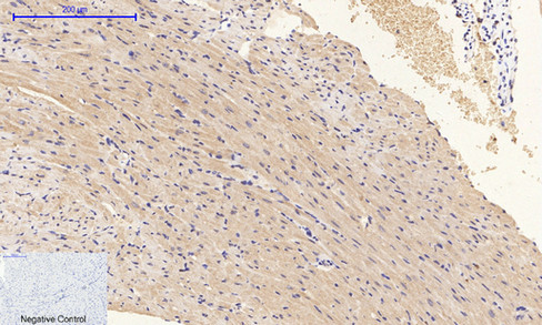
Immunohistochemical analysis of paraffin-embedded Mouse-heart tissue. 1,PI 3-kinase p85/p55 (phospho Tyr467/199) Polyclonal Antibody was diluted at 1:200(4°C,overnight). 2, Sodium citrate pH 6.0 was used for antibody retrieval(>98°C,20min). 3,Secondary antibody was diluted at 1:200(room tempeRature, 30min). Negative control was used by secondary antibody only.IHC
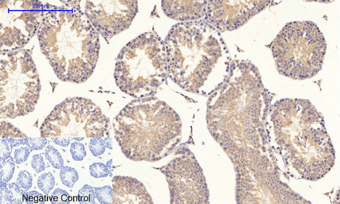
Immunohistochemical analysis of paraffin-embedded Mouse-testis tissue. 1,PI 3-kinase p85/p55 (phospho Tyr467/199) Polyclonal Antibody was diluted at 1:200(4°C,overnight). 2, Sodium citrate pH 6.0 was used for antibody retrieval(>98°C,20min). 3,Secondary antibody was diluted at 1:200(room tempeRature, 30min). Negative control was used by secondary antibody only.ICC/IF
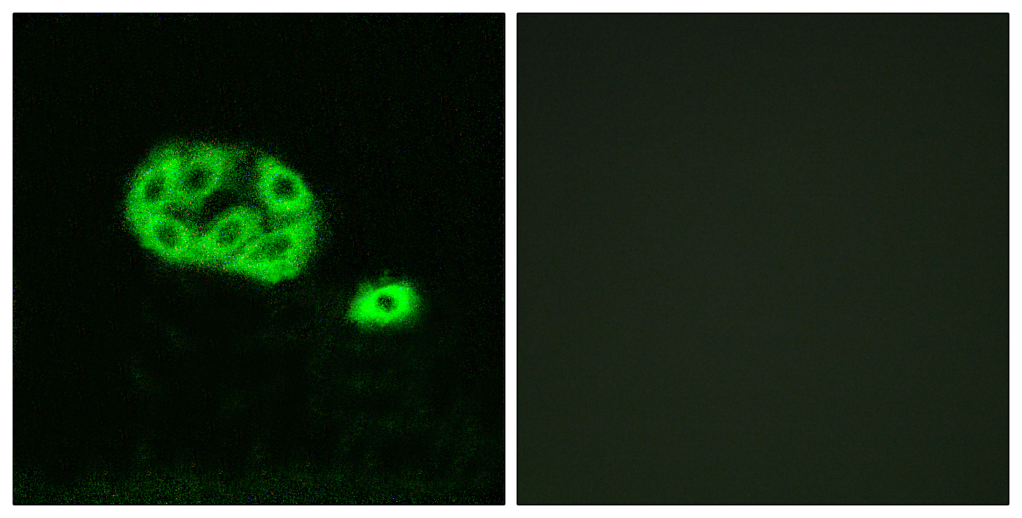
Immunofluorescence analysis of NIH/3T3 cells, using PI3-kinase p85-alpha/gamma (Phospho-Tyr467/199) Antibody. The picture on the right is blocked with the phospho peptide.IF-P
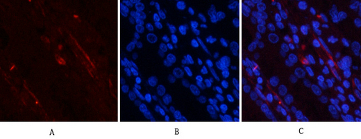
Immunofluorescence analysis of human-stomach tissue. 1,PI 3-kinase p85/p55 (phospho Tyr467/199) Polyclonal Antibody(red) was diluted at 1:200(4°C,overnight). 2, Cy3 labled Secondary antibody was diluted at 1:300(room temperature, 50min).3, Picture B: DAPI(blue) 10min. Picture A:Target. Picture B: DAPI. Picture C: merge of A+BIF-P
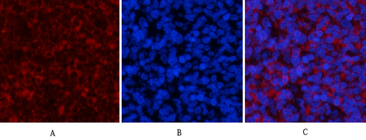
Immunofluorescence analysis of rat-spleen tissue. 1,PI 3-kinase p85/p55 (phospho Tyr467/199) Polyclonal Antibody(red) was diluted at 1:200(4°C,overnight). 2, Cy3 labled Secondary antibody was diluted at 1:300(room temperature, 50min).3, Picture B: DAPI(blue) 10min. Picture A:Target. Picture B: DAPI. Picture C: merge of A+B| Product Name | PI 3 kinase p85/p55 (Phospho Tyr467/199) Rabbit pAb |
|---|---|
| Antibody Type | Primary Antibodies |
| Immunogen | The antiserum was produced against synthesized peptide derived from human PI3-kinase p85-alpha/gamma around the phosphorylation site of Tyr467/199. AA range:436-485 |
| Modification | p-Tyr467/199 |
| Clonality | Polyclonal |
|---|---|
| Isotype | IgG |
| Host Species | Rabbit |
| Tested Applications | ICC/IFIF-PIHCWB |
| WB:1:500-1:2000 IHC:100-1:300 ICC/IF:1:200-1:1000 IF-P:1:200-1:1000 |
|
| Species Reactivity | HumanMonkeyMousePigRat |
| Concentration | 1mg/ml |
| Purification | Affinity purified |
| Gene Symbol | PIK3R1,PIK3R3 |
|---|---|
| Gene Synonyms | p85 AGM7 GRB1 IMD36 p85alpha p85-ALPHA p55 p55PIK p55-GAMMA |
| Gene Full Name | Phosphatidylinositol 3-kinase regulatory subunit alpha/gamma |
| Gene Summary | Phosphatidylinositol 3-kinase phosphorylates the inositol ring of phosphatidylinositol at the 3-prime position. The enzyme comprises a 110 kD catalytic subunit and a regulatory subunit of either 85, 55, or 50 kD. This gene encodes the 85 kD regulatory subunit. Phosphatidylinositol 3-kinase plays an important role in the metabolic actions of insulin, and a mutation in this gene has been associated with insulin resistance. Alternative splicing of this gene results in four transcript variants encoding different isoforms. [provided by RefSeq, Jun 2011] |
| Alternative Names | PIK3R1 GRB1 Phosphatidylinositol 3-kinase regulatory subunit alpha PI3-kinase regulatory subunit alpha PI3K regulatory subunit alpha PtdIns-3-kinase regulatory subunit alpha Phosphatidylinositol 3-kinase 85 kDa regulatory subunit alpha PI3-kinase subunit p85-alpha PtdIns-3-kinase regulatory subunit p85-alpha |
| Molecular Weight(MW) | 55kDa,85kDa |
| Cellular Localization | Cell-cell junction, cis-Golgi network, cytoplasm, cytosol, membrane, nucleus. |
WB
Western Blot analysis of various cells using primary antibody diluted at 1:1000(4°C overnight). Secondary antibody:Goat Anti-rabbit IgG IRDye 800( diluted at 1:5000, 25°C, 1 hour)
WB
Western blot analysis of lysates from COS7 cells treated with H2O2 100uM 30', using PI3-kinase p85-alpha/gamma (Phospho-Tyr467/199) Antibody. The lane on the right is blocked with the phospho peptide.
IHC
Immunohistochemical analysis of paraffin-embedded Human-colon tissue. 1,PI 3-kinase p85/p55 (phospho Tyr467/199) Polyclonal Antibody was diluted at 1:200(4°C,overnight). 2, Sodium citrate pH 6.0 was used for antibody retrieval(>98°C,20min). 3,Secondary antibody was diluted at 1:200(room tempeRature, 30min). Negative control was used by secondary antibody only.
IHC
Immunohistochemical analysis of paraffin-embedded Human-liver tissue. 1,PI 3-kinase p85/p55 (phospho Tyr467/199) Polyclonal Antibody was diluted at 1:200(4°C,overnight). 2, Sodium citrate pH 6.0 was used for antibody retrieval(>98°C,20min). 3,Secondary antibody was diluted at 1:200(room tempeRature, 30min). Negative control was used by secondary antibody only.
IHC
Immunohistochemical analysis of paraffin-embedded Rat-liver tissue. 1,PI 3-kinase p85/p55 (phospho Tyr467/199) Polyclonal Antibody was diluted at 1:200(4°C,overnight). 2, Sodium citrate pH 6.0 was used for antibody retrieval(>98°C,20min). 3,Secondary antibody was diluted at 1:200(room tempeRature, 30min). Negative control was used by secondary antibody only.
IHC
Immunohistochemical analysis of paraffin-embedded Mouse-heart tissue. 1,PI 3-kinase p85/p55 (phospho Tyr467/199) Polyclonal Antibody was diluted at 1:200(4°C,overnight). 2, Sodium citrate pH 6.0 was used for antibody retrieval(>98°C,20min). 3,Secondary antibody was diluted at 1:200(room tempeRature, 30min). Negative control was used by secondary antibody only.
IHC
Immunohistochemical analysis of paraffin-embedded Mouse-testis tissue. 1,PI 3-kinase p85/p55 (phospho Tyr467/199) Polyclonal Antibody was diluted at 1:200(4°C,overnight). 2, Sodium citrate pH 6.0 was used for antibody retrieval(>98°C,20min). 3,Secondary antibody was diluted at 1:200(room tempeRature, 30min). Negative control was used by secondary antibody only.
ICC/IF
Immunofluorescence analysis of NIH/3T3 cells, using PI3-kinase p85-alpha/gamma (Phospho-Tyr467/199) Antibody. The picture on the right is blocked with the phospho peptide.
IF-P
Immunofluorescence analysis of human-stomach tissue. 1,PI 3-kinase p85/p55 (phospho Tyr467/199) Polyclonal Antibody(red) was diluted at 1:200(4°C,overnight). 2, Cy3 labled Secondary antibody was diluted at 1:300(room temperature, 50min).3, Picture B: DAPI(blue) 10min. Picture A:Target. Picture B: DAPI. Picture C: merge of A+B
IF-P
Immunofluorescence analysis of rat-spleen tissue. 1,PI 3-kinase p85/p55 (phospho Tyr467/199) Polyclonal Antibody(red) was diluted at 1:200(4°C,overnight). 2, Cy3 labled Secondary antibody was diluted at 1:300(room temperature, 50min).3, Picture B: DAPI(blue) 10min. Picture A:Target. Picture B: DAPI. Picture C: merge of A+B| Application Notes | WB:1:500-1:2000 IHC:100-1:300 ICC/IF:1:200-1:1000 IF-P:1:200-1:1000 |
|---|
| Form | Liquid |
|---|---|
| Storage Instructions | -15°C to -25°C/1 year(Do not lower than -25°C) |
| Storage Buffer | Liquid in PBS containing 50% glycerol, 0.5% BSA and 0.02% sodium azide. |


