WB
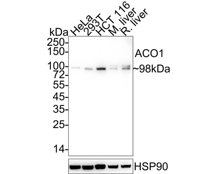
Western blot analysis of ACO1 on different lysates with Rabbit anti-ACO1 antibody at 1/2,000 dilution. Lane 1: HeLa cell lysate (20 µg/Lane), Lane 2: 293T cell lysate (20 µg/Lane), Lane 3: HCT 116 cell lysate (20 µg/Lane), Lane 4: Mouse liver tissue lysate (40 µg/Lane), Lane 5: Rat liver tissue lysate (40 µg/Lane), Exposure time: 5 minutes 10 seconds; 4-20% SDS-PAGE gel. Proteins were transferred to a PVDF membrane and blocked with 5% NFDM/TBST for 1 hour at room temperature. The primary antibody at 1/2,000 dilution was used in 5% NFDM/TBST at 4℃ overnight. Goat Anti-Rabbit IgG - HRP Secondary Antibody at 1/50,000 dilution was used for 1 hour at room temperature.FC
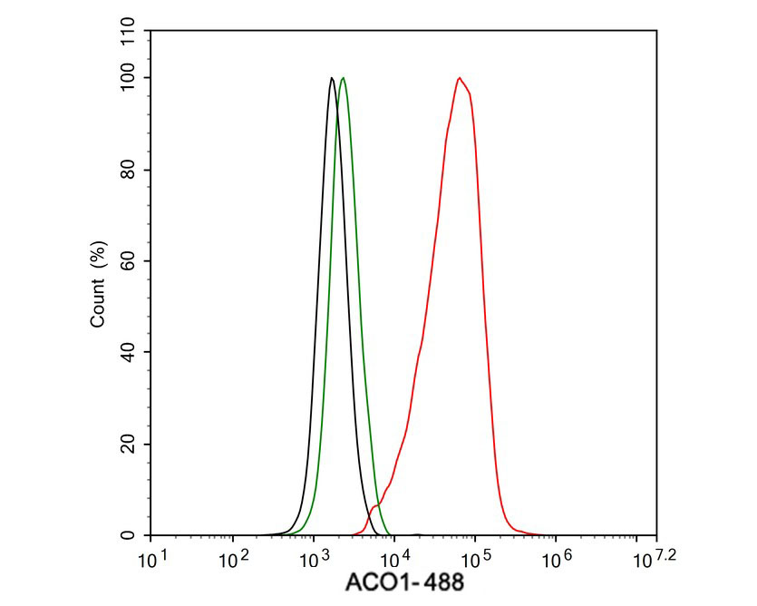
Flow cytometric analysis of HeLa cells labeling ACO1. Cells were fixed and permeabilized. Then stained with the primary antibody (1μg/mL) (red) compared with Rabbit IgG Isotype Control (green). After incubation of the primary antibody at +4℃ for an hour, the cells were stained with a 488 conjugate-Goat anti-Rabbit IgG Secondary antibody at 1/1,000 dilution for 30 minutes at +4℃. Unlabelled sample was used as a control (cells without incubation with primary antibody; black).| Product Name | ACO1 Recombinant Rabbit Monoclonal Antibody |
|---|---|
| Antibody Type | Primary Antibodies |
| Immunogen | Synthetic peptide within Human ACO1 aa 1-50 / 889. |
| Clonality | Monoclonal |
|---|---|
| Isotype | IgG |
| Host Species | Rabbit |
| Tested Applications | FCWB |
| WB:1:2000-1:5000 FC:1:1000 |
|
| Species Reactivity | HumanMouseRat |
| Concentration | 1mg/ml |
| Purification | Protein A |
| Gene Symbol | ACO1 |
|---|---|
| Gene Synonyms | IRP1 ACONS HEL60 IREB1 IREBP IREBP1 |
| Gene Full Name | aconitase 1 |
| Gene Summary | The protein encoded by this gene is a bifunctional, cytosolic protein that functions as an essential enzyme in the TCA cycle and interacts with mRNA to control the levels of iron inside cells. When cellular iron levels are high, this protein binds to a 4Fe-4S cluster and functions as an aconitase. Aconitases are iron-sulfur proteins that function to catalyze the conversion of citrate to isocitrate. When cellular iron levels are low, the protein binds to iron-responsive elements (IREs), which are stem-loop structures found in the 5' UTR of ferritin mRNA, and in the 3' UTR of transferrin receptor mRNA. When the protein binds to IRE, it results in repression of translation of ferritin mRNA, and inhibition of degradation of the otherwise rapidly degraded transferrin receptor mRNA. The encoded protein has been identified as a moonlighting protein based on its ability to perform mechanistically distinct functions. Alternative splicing results in multiple transcript variants [provided by RefSeq, Jan 2014] |
| Alternative Names | ACO 1 antibody ACO1 antibody ACOC_HUMAN antibody Aconitase 1 soluble antibody Aconitase antibody Aconitase1 antibody Aconitate hydratase antibody ACONS antibody Citrate hydro lyase antibody Citrate hydro-lyase antibody ACO 1 antibody ACO1 antibody ACOC_HUMAN antibody Aconitase 1 soluble antibody Aconitase antibody Aconitase1 antibody Aconitate hydratase antibody ACONS antibody Citrate hydro lyase antibody Citrate hydro-lyase antibody Cytoplasmic aconitate hydratase antibody Ferritin repressor protein antibody IRE BP 1 antibody IRE-BP 1 antibody IREB 1 antibody IREB1 antibody IREBP antibody IREBP1 antibody Iron regulatory protein 1 antibody Iron responsive element binding protein 1 antibody Iron-responsive element-binding protein 1 antibody IRP 1 antibody IRP1 antibody OTTHUMP00000045233 antibody |
| Molecular Weight(MW) | 98 kDa |
| Cellular Localization | Cytoplasm, cytosol. |
WB
Western blot analysis of ACO1 on different lysates with Rabbit anti-ACO1 antibody at 1/2,000 dilution. Lane 1: HeLa cell lysate (20 µg/Lane), Lane 2: 293T cell lysate (20 µg/Lane), Lane 3: HCT 116 cell lysate (20 µg/Lane), Lane 4: Mouse liver tissue lysate (40 µg/Lane), Lane 5: Rat liver tissue lysate (40 µg/Lane), Exposure time: 5 minutes 10 seconds; 4-20% SDS-PAGE gel. Proteins were transferred to a PVDF membrane and blocked with 5% NFDM/TBST for 1 hour at room temperature. The primary antibody at 1/2,000 dilution was used in 5% NFDM/TBST at 4℃ overnight. Goat Anti-Rabbit IgG - HRP Secondary Antibody at 1/50,000 dilution was used for 1 hour at room temperature.
FC
Flow cytometric analysis of HeLa cells labeling ACO1. Cells were fixed and permeabilized. Then stained with the primary antibody (1μg/mL) (red) compared with Rabbit IgG Isotype Control (green). After incubation of the primary antibody at +4℃ for an hour, the cells were stained with a 488 conjugate-Goat anti-Rabbit IgG Secondary antibody at 1/1,000 dilution for 30 minutes at +4℃. Unlabelled sample was used as a control (cells without incubation with primary antibody; black).| Application Notes | WB:1:2000-1:5000 FC:1:1000 |
|---|
| Form | Liquid |
|---|---|
| Storage Instructions | Store at +4℃ after thawing. Aliquot store at -20℃. Avoid repeated freeze / thaw cycles. |
| Storage Buffer | 1*TBS (pH7.4), 0.05% BSA, 40% Glycerol. Preservative: 0.05% Sodium Azide. |


