WB
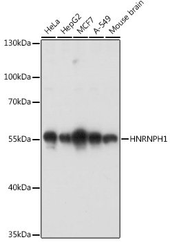
Western blot analysis of various lysates using HNRNPH1 Rabbit pAb at 1:3000 dilution. Secondary antibody: HRP-conjugated Goat anti-Rabbit IgG (H+L) at 1:10000 dilution. Lysates/proteins: 25μg per lane. Blocking buffer: 3% nonfat dry milk in TBST. Detection: ECL Basic Kit. Exposure time: 90s.WB
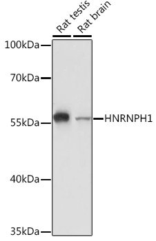
Western blot analysis of various lysates using HNRNPH1 Rabbit pAb at 1:3000 dilution. Secondary antibody: HRP-conjugated Goat anti-Rabbit IgG (H+L) at 1:10000 dilution. Lysates/proteins: 25μg per lane. Blocking buffer: 3% nonfat dry milk in TBST. Detection: ECL Basic Kit. Exposure time: 90s.IHC
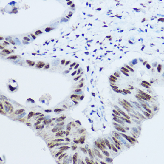
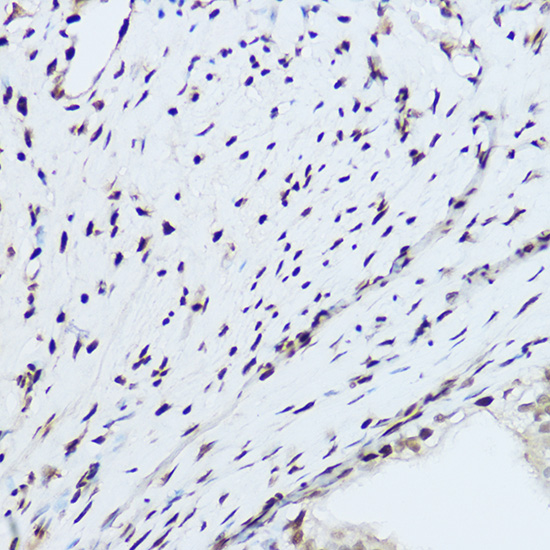
Immunohistochemistry analysis of paraffin embedded Human colon carcinoma using HNRNPH1 Rabbit pAb at dilution of 1:100 (40x lens). High pressure antigen retrieval performed with 0.01M Citrate buffer (pH 6.0) prior to IHC staining.IHC
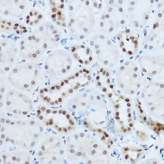
Immunohistochemistry analysis of paraffin embedded Mouse kidney using HNRNPH1 Rabbit pAb at dilution of 1:100 (40x lens). High pressure antigen retrieval performed with 0.01M Citrate buffer (pH 6.0) prior to IHC staining.IHC
Immunohistochemistry analysis of paraffin embedded Rat ovary using HNRNPH1 Rabbit pAb at dilution of 1:100 (40x lens). High pressure antigen retrieval performed with 0.01M Citrate buffer (pH 6.0) prior to IHC staining.ICC/IF
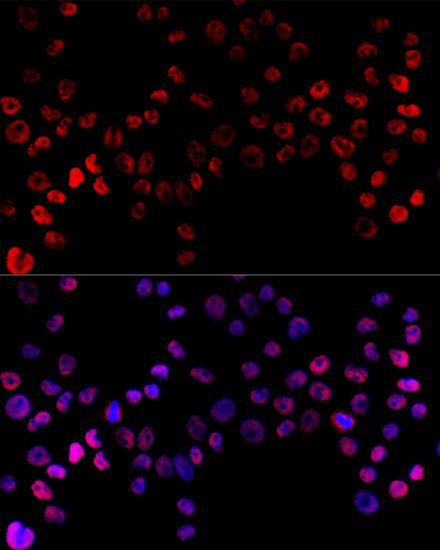
Immunofluorescence analysis of 293T cells using HNRNPH1 Rabbit pAb at dilution of 1:100 (40x lens). Secondary antibody: Cy3-conjugated Goat anti-Rabbit IgG (H+L) at 1:500 dilution. Blue: DAPI for nuclear staining.| Product Name | HNRNPH1 Rabbit pAb |
|---|---|
| Antibody Type | Primary Antibodies |
| Immunogen | Recombinant protein. |
| Clonality | Polyclonal |
|---|---|
| Isotype | IgG |
| Host Species | Rabbit |
| Tested Applications | ICC/IFIHCWB |
| WB:1:1000-1:5000 IHC:1:50-1:200 ICC/IF:1:50-1:200 |
|
| Species Reactivity | HumanMouseRat |
| Concentration | 1mg/ml |
| Purification | Affinity purified |
| Gene Symbol | HNRNPH1 |
|---|---|
| Gene Synonyms | HNRPH HNRPH1 NEDCDS hnRNPH |
| Gene Full Name | heterogeneous nuclear ribonucleoprotein H1 |
| Gene Summary | This gene encodes a member of a subfamily of ubiquitously expressed heterogeneous nuclear ribonucleoproteins (hnRNPs). The hnRNPs are RNA binding proteins that complex with heterogeneous nuclear RNA. These proteins are associated with pre-mRNAs in the nucleus and appear to influence pre-mRNA processing and other aspects of mRNA metabolism and transport. While all of the hnRNPs are present in the nucleus, some may shuttle between the nucleus and the cytoplasm. The hnRNP proteins have distinct nucleic acid binding properties. The protein encoded by this gene has three repeats of quasi-RRM domains that bind to RNA and is very similar to the family member HNRPF. This gene may be associated with hereditary lymphedema type I. Alternatively spliced transcript variants have been described [provided by RefSeq, Mar 2012] |
| Alternative Names | HNRPH HNRPH1 NEDCDS hnRNPH HNRNPH1 |
| Molecular Weight(MW) | 49kDa(Observed MW 55kDa) |
| Cellular Localization | Nucleus, nucleoplasm. |
WB
Western blot analysis of various lysates using HNRNPH1 Rabbit pAb at 1:3000 dilution. Secondary antibody: HRP-conjugated Goat anti-Rabbit IgG (H+L) at 1:10000 dilution. Lysates/proteins: 25μg per lane. Blocking buffer: 3% nonfat dry milk in TBST. Detection: ECL Basic Kit. Exposure time: 90s.
WB
Western blot analysis of various lysates using HNRNPH1 Rabbit pAb at 1:3000 dilution. Secondary antibody: HRP-conjugated Goat anti-Rabbit IgG (H+L) at 1:10000 dilution. Lysates/proteins: 25μg per lane. Blocking buffer: 3% nonfat dry milk in TBST. Detection: ECL Basic Kit. Exposure time: 90s.
IHC
Immunohistochemistry analysis of paraffin embedded Human colon carcinoma using HNRNPH1 Rabbit pAb at dilution of 1:100 (40x lens). High pressure antigen retrieval performed with 0.01M Citrate buffer (pH 6.0) prior to IHC staining.
IHC
Immunohistochemistry analysis of paraffin embedded Mouse kidney using HNRNPH1 Rabbit pAb at dilution of 1:100 (40x lens). High pressure antigen retrieval performed with 0.01M Citrate buffer (pH 6.0) prior to IHC staining.
IHC
Immunohistochemistry analysis of paraffin embedded Rat ovary using HNRNPH1 Rabbit pAb at dilution of 1:100 (40x lens). High pressure antigen retrieval performed with 0.01M Citrate buffer (pH 6.0) prior to IHC staining.
ICC/IF
Immunofluorescence analysis of 293T cells using HNRNPH1 Rabbit pAb at dilution of 1:100 (40x lens). Secondary antibody: Cy3-conjugated Goat anti-Rabbit IgG (H+L) at 1:500 dilution. Blue: DAPI for nuclear staining.| Application Notes | WB:1:1000-1:5000 IHC:1:50-1:200 ICC/IF:1:50-1:200 |
|---|
| Form | Liquid |
|---|---|
| Storage Instructions | Store at -20℃. Avoid freeze / thaw cycles. |
| Storage Buffer | Buffer: PBS with 0.05% proclin300, 0.05% BSA, 50% glycerol, pH7.3. |


