WB
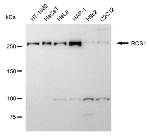
Western blotting analysis using ROS1 antibody. Total cell lysates (30 μg) from various cell lines were loaded and separated by SDS-PAGE. The blot was incubated with ROS1 antibody and HRP-conjugated goat anti-mouse secondary antibody respectively.FC
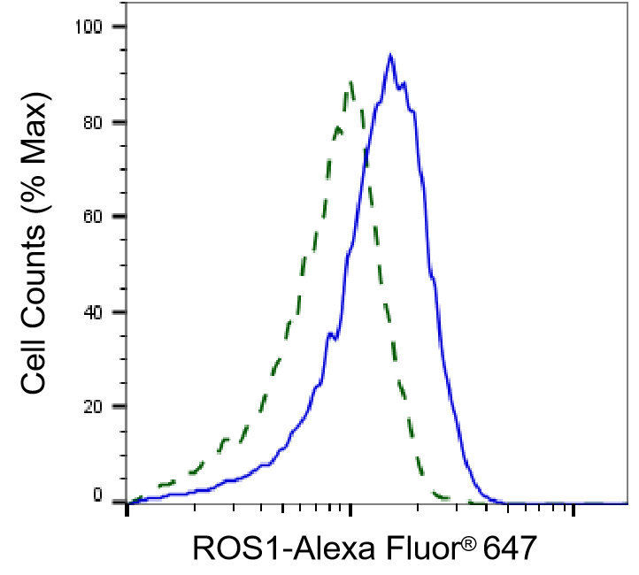
Flow cytometric analysis of ROS1 expression in HAP-1 cells using ROS1 antibody. Green, isotype control; red, ROS1.| Product Name | Mouse anti-ROS1 Monoclonal antibody |
|---|---|
| Antibody Type | Primary Antibodies |
| Immunogen | Recombinant Human ROS1 protein |
| Clonality | Monoclonal |
|---|---|
| Isotype | IgG2a |
| Host Species | Mouse |
| Tested Applications | FCWB |
| WB:1:100-1:500 FC:1:200-1:2000 |
|
| Species Reactivity | HumanMouseRat |
| Concentration | 1mg/ml |
| Purification | Affinity purified |
| Gene Symbol | ROS1 |
|---|---|
| Gene Synonyms | ROS MCF3 c-ros-1 |
| Gene Full Name | ROS proto-oncogene 1, receptor tyrosine kinase |
| Gene Summary | This proto-oncogene, highly-expressed in a variety of tumor cell lines, belongs to the sevenless subfamily of tyrosine kinase insulin receptor genes. The protein encoded by this gene is a type I integral membrane protein with tyrosine kinase activity. The protein may function as a growth or differentiation factor receptor. [provided by RefSeq, Jul 2008] |
| Alternative Names | ROS1 ROS Proto-Oncogene 1 Receptor Tyrosine Kinase MCF3 ROS C-Ros-1 V-Ros Avian UR2 Sarcoma Virus Oncogene Homolog 1 C-Ros Oncogene 1 Receptor Tyrosine Kinase Proto-Oncogene Tyrosine-Protein Kinase ROS Proto-Oncogene C-Ros-1 V-Ros UR2 Sarcoma Virus Oncogene Homolog 1 (Avian) ROS Proto-Oncogene 1 Receptor Tyrosine Kinase Transmembrane Tyrosine-Specific Protein Kinase Receptor Tyrosine Kinase C-Ros Oncogene 1 C-Ros Receptor Tyrosine Kinase Proto-Oncogene C-Ros EC 2.7.10.1 |
| Cellular Localization | Cell membrane; Single-pass type I membrane protein. |
WB
Western blotting analysis using ROS1 antibody. Total cell lysates (30 μg) from various cell lines were loaded and separated by SDS-PAGE. The blot was incubated with ROS1 antibody and HRP-conjugated goat anti-mouse secondary antibody respectively.
FC
Flow cytometric analysis of ROS1 expression in HAP-1 cells using ROS1 antibody. Green, isotype control; red, ROS1.| Application Notes | WB:1:100-1:500 FC:1:200-1:2000 |
|---|
| Form | Liquid |
|---|---|
| Storage Instructions | Upon receipt, store at -20°C or -80°C. Avoid repeated freeze. |
| Storage Buffer | 0.03% Proclin 300 Constituents: 50% Glycerol, 0.01M PBS, PH 7.4 |


