WB
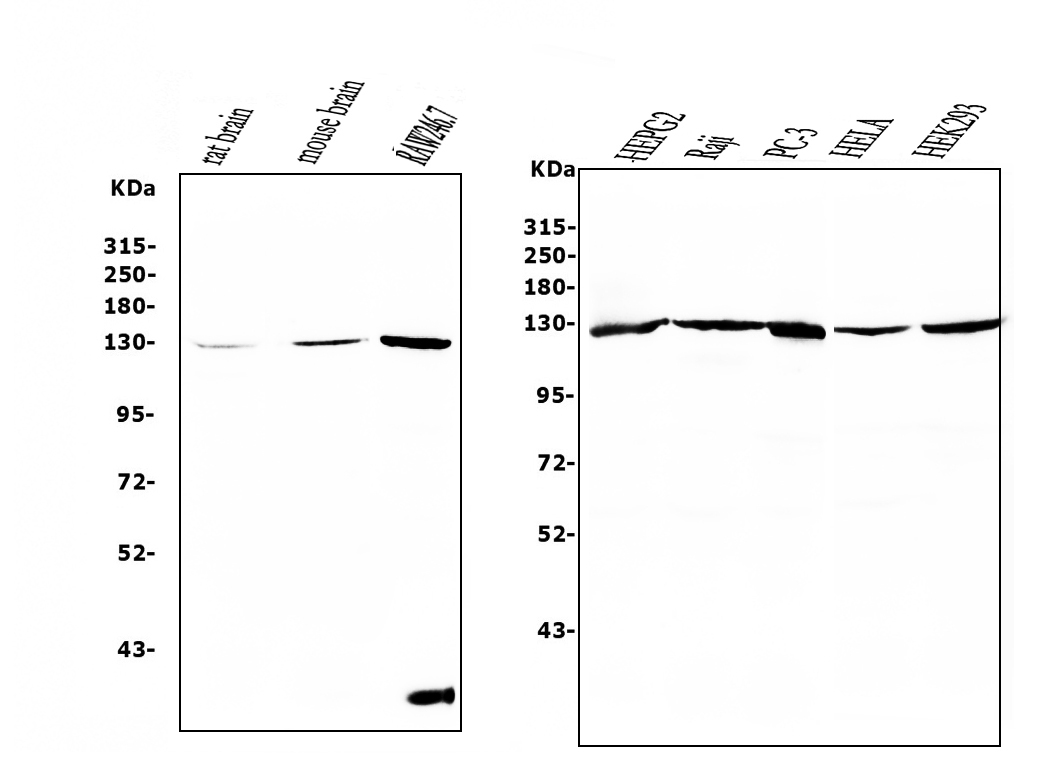
Western blot analysis of UPF1 using anti-UPF1 antibody. The sample well of each lane was loaded with 30 ug of sample under reducing conditions. After electrophoresis, proteins were transferred to a membrane. Then the membrane was incubated with mouse anti-UPF1 antigen affinity purified monoclonal antibody at a dilution of 1:1000 and probed with a goat anti-mouse IgG-HRP secondary antibody. The signal is developed using ECL Plus Western Blotting Substrate.IHC
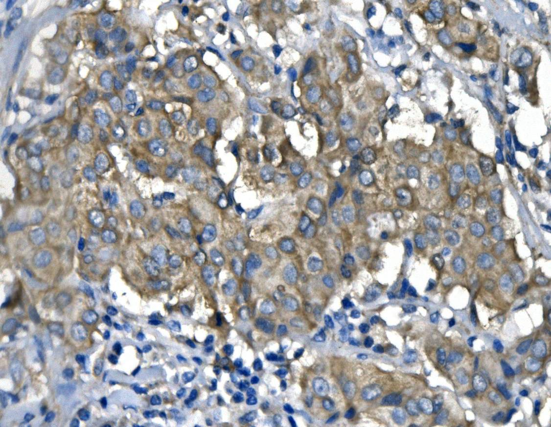
IHC analysis of UPF1 using anti-UPF1 antibody. UPF1 was detected in a paraffin-embedded section of human mammary cancer tissue. Biotinylated goat anti-mouse IgG was used as secondary antibody. The tissue section was incubated with mouse anti-UPF1 Antibody at a dilution of 1:200 and developed using Strepavidin-Biotin-Complex (SABC) with DAB as the chromogen.IHC
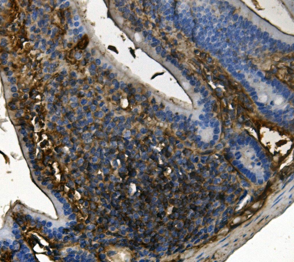
IHC analysis of UPF1 using anti-UPF1 antibody. UPF1 was detected in a paraffin-embedded section of mouse intestine tissue. Biotinylated goat anti-mouse IgG was used as secondary antibody. The tissue section was incubated with mouse anti-UPF1 Antibody at a dilution of 1:200 and developed using Strepavidin-Biotin-Complex (SABC) with DAB as the chromogen.IHC
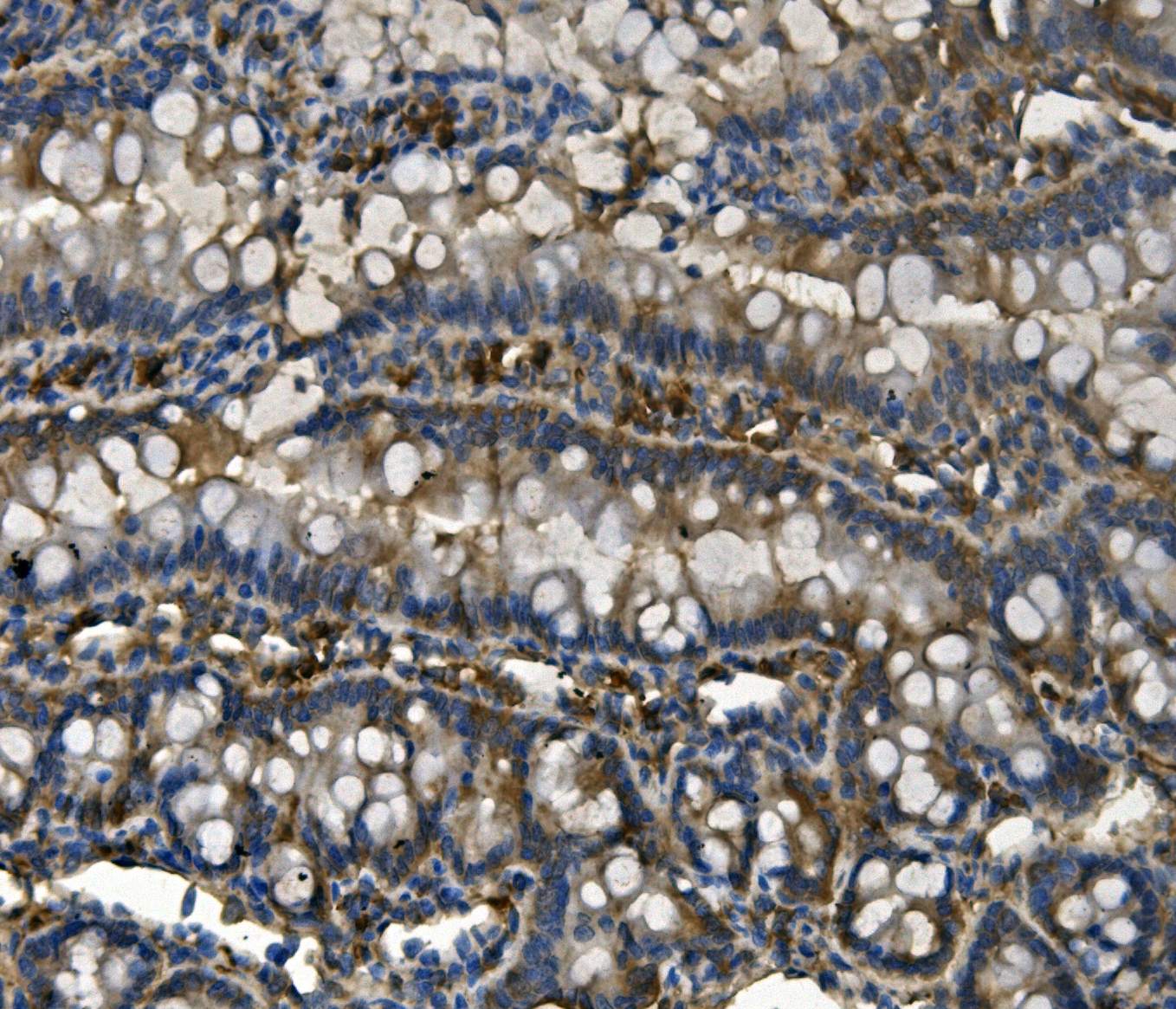
IHC analysis of UPF1 using anti-UPF1 antibody. UPF1 was detected in a paraffin-embedded section of rat intestine tissue. Biotinylated goat anti-mouse IgG was used as secondary antibody. The tissue section was incubated with mouse anti-UPF1 Antibody (M00900) at a dilution of 1:200 and developed using Strepavidin-Biotin-Complex (SABC) with DAB as the chromogen.ICC/IF
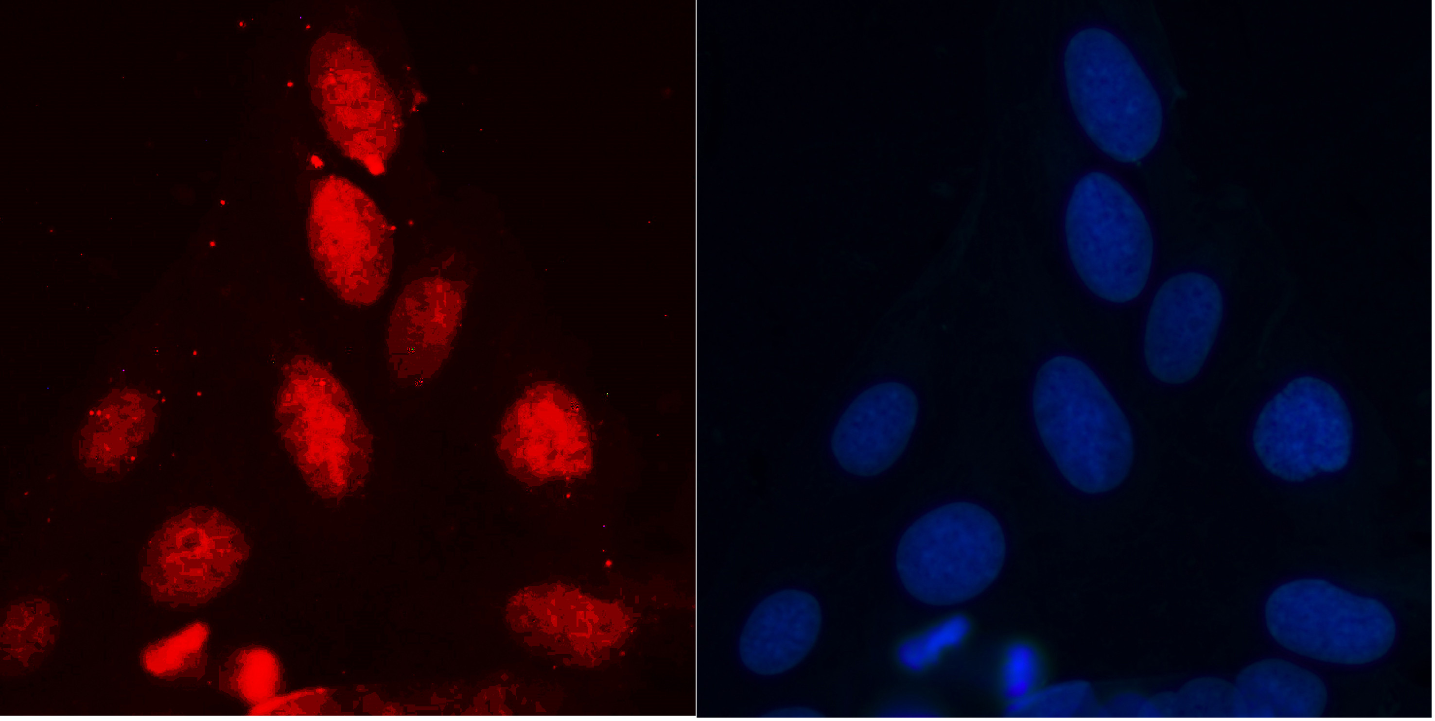
ICC/IF analysis of UPF1 using anti-UPF1 antibody. UPF1 was detected in an immunocytochemical section of U2OS cells. The section was incubated with mouse anti-UPF1 Antibody at a dilution of 1:100. Fluoro594-conjugated Anti-mouse IgG Secondary Antibody (red) was used as secondary antibody. The section was counterstained with DAPI(Blue).| Product Name | Mouse monoclonal antibody to UPF1 |
|---|---|
| Antibody Type | Primary Antibodies |
| Immunogen | A synthetic peptide corresponding to a sequence in the middle region of human RENT1/hUPF1, identical to the related mouse and rat sequences. |
| Clonality | monoclonal |
|---|---|
| Isotype | IgG2b |
| Host Species | Mouse |
| Tested Applications | ICC/IFIHCWB |
| WB:1:500-1:2000 IHC:1:50-1:400 ICC/IF:1:50-1:400 |
|
| Species Reactivity | HumanMouseRat |
| Concentration | 0.5mg/ml |
| Purification | Protein G |
| Gene Symbol | UPF1 |
|---|---|
| Gene Synonyms | UTF HUPF1 NORF1 RENT1 smg-2 pNORF1 |
| Gene Full Name | UPF1 RNA helicase and ATPase |
| Gene Summary | This gene encodes a protein that is part of a post-splicing multiprotein complex involved in both mRNA nuclear export and mRNA surveillance. mRNA surveillance detects exported mRNAs with truncated open reading frames and initiates nonsense-mediated mRNA decay (NMD). When translation ends upstream from the last exon-exon junction, this triggers NMD to degrade mRNAs containing premature stop codons. This protein is located only in the cytoplasm. When translation ends, it interacts with the protein that is a functional homolog of yeast Upf2p to trigger mRNA decapping. Use of multiple polyadenylation sites has been noted for this gene. Alternative splicing results in multiple transcript variants. [provided by RefSeq, Jul 2014] |
| Alternative Names | ATP dependent helicase RENT1 FLJ43809 FLJ46894 HUPF1 KIAA0221 NORF1 pNORF1 RENT1 UPF1 |
| Molecular Weight(MW) | 124 kDa(Observed band size: 130 kDa) |
| Cellular Localization | Cytoplasm,Nucleus |
WB
Western blot analysis of UPF1 using anti-UPF1 antibody. The sample well of each lane was loaded with 30 ug of sample under reducing conditions. After electrophoresis, proteins were transferred to a membrane. Then the membrane was incubated with mouse anti-UPF1 antigen affinity purified monoclonal antibody at a dilution of 1:1000 and probed with a goat anti-mouse IgG-HRP secondary antibody. The signal is developed using ECL Plus Western Blotting Substrate.
IHC
IHC analysis of UPF1 using anti-UPF1 antibody. UPF1 was detected in a paraffin-embedded section of human mammary cancer tissue. Biotinylated goat anti-mouse IgG was used as secondary antibody. The tissue section was incubated with mouse anti-UPF1 Antibody at a dilution of 1:200 and developed using Strepavidin-Biotin-Complex (SABC) with DAB as the chromogen.
IHC
IHC analysis of UPF1 using anti-UPF1 antibody. UPF1 was detected in a paraffin-embedded section of mouse intestine tissue. Biotinylated goat anti-mouse IgG was used as secondary antibody. The tissue section was incubated with mouse anti-UPF1 Antibody at a dilution of 1:200 and developed using Strepavidin-Biotin-Complex (SABC) with DAB as the chromogen.
IHC
IHC analysis of UPF1 using anti-UPF1 antibody. UPF1 was detected in a paraffin-embedded section of rat intestine tissue. Biotinylated goat anti-mouse IgG was used as secondary antibody. The tissue section was incubated with mouse anti-UPF1 Antibody (M00900) at a dilution of 1:200 and developed using Strepavidin-Biotin-Complex (SABC) with DAB as the chromogen.
ICC/IF
ICC/IF analysis of UPF1 using anti-UPF1 antibody. UPF1 was detected in an immunocytochemical section of U2OS cells. The section was incubated with mouse anti-UPF1 Antibody at a dilution of 1:100. Fluoro594-conjugated Anti-mouse IgG Secondary Antibody (red) was used as secondary antibody. The section was counterstained with DAPI(Blue).| Application Notes | WB:1:500-1:2000 IHC:1:50-1:400 ICC/IF:1:50-1:400 |
|---|
| Form | Liquid |
|---|---|
| Storage Instructions | 12 months from date of receipt,-20℃ as supplied. 6 months 2 to 8℃ after reconstitution. Avoid repeated freezing and thawing. |
| Storage Buffer | 500 ug/ml antibody with PBS, 0.02% NaN3, 1 mg/ml BSA and 50% glycerol. |


