WB
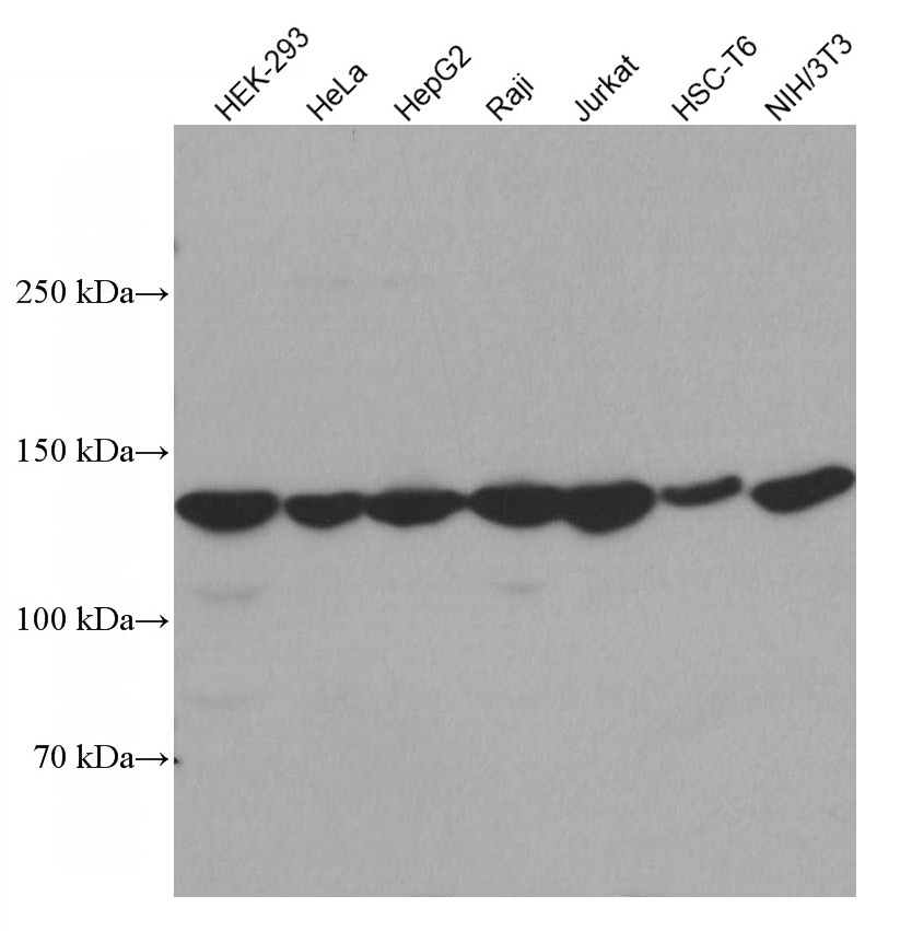
Various lysates were subjected to SDS PAGE followed by western blot with UPF1 antibody at dilution of 1:5000 incubated at room temperature for 1.5 hours.WB
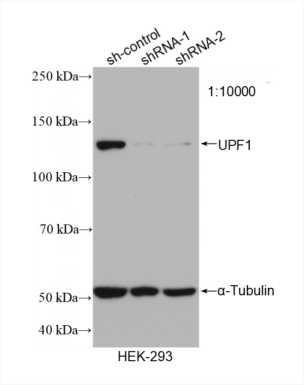
WB result of UPF1 antibody (1:10000; incubated at room temperature for 1.5 hours) with sh-Control and sh-UPF1 transfected HEK-293 cells.WB
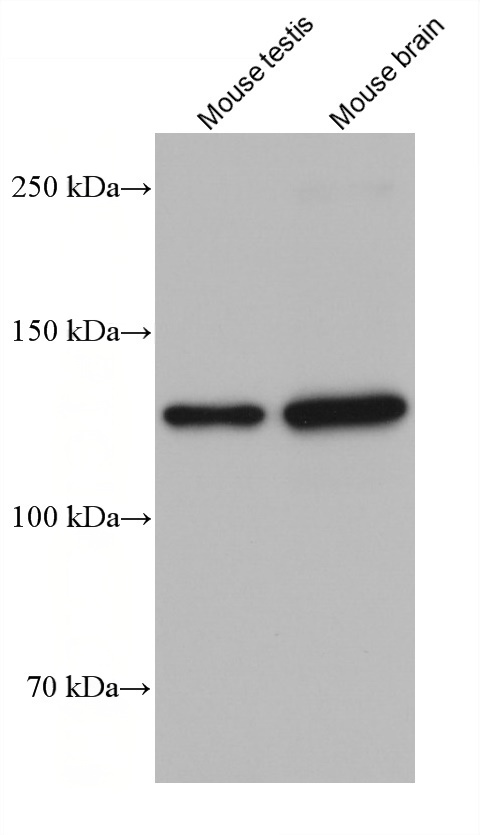
Various lysates were subjected to SDS PAGE followed by western blot with UPF1 antibody at dilution of 1:5000 incubated at room temperature for 1.5 hours.IHC
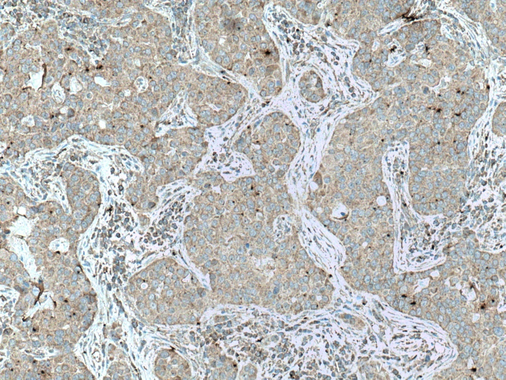
Immunohistochemical analysis of paraffin-embedded human breast cancer tissue slide using UPF1 antibody at dilution of 1:500 (under 10x lens. Heat mediated antigen retrieval with Tris-EDTA buffer (pH 9.0).IHC
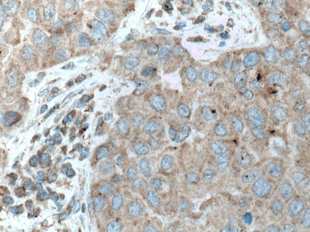
Immunohistochemical analysis of paraffin-embedded human breast cancer tissue slide using UPF1 antibody at dilution of 1:500 (under 40x lens. Heat mediated antigen retrieval with Tris-EDTA buffer (pH 9.0).ICC/IF
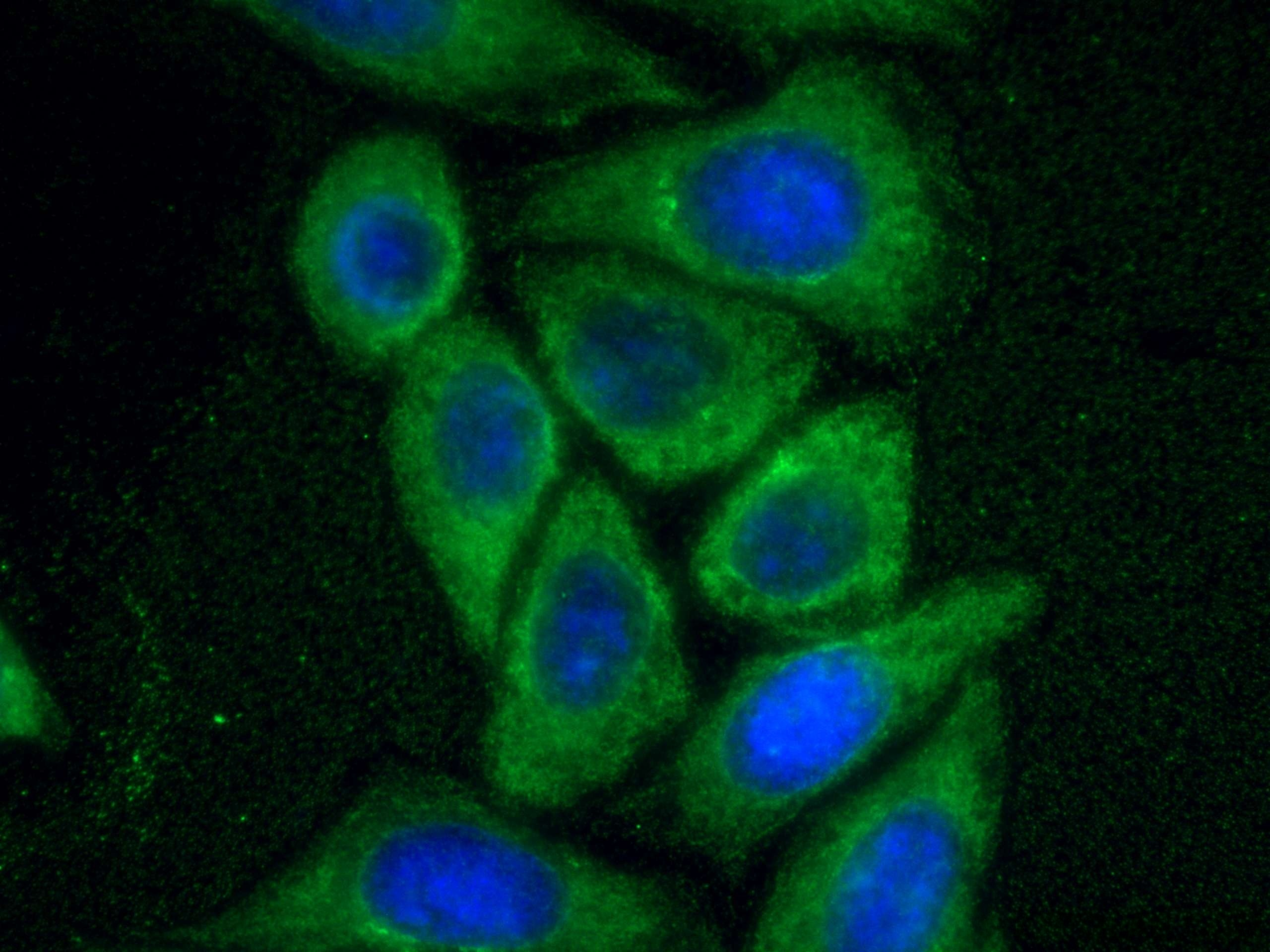
Immunofluorescent analysis of (-20°C Methanol) fixed HepG2 cells using UPF1 antibody at dilution of 1:800 and CoraLite®488-Conjugated Goat Anti-Mouse IgG(H+L).FC
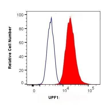
1X10^6 HepG2 cells were intracellularly stained with 0.4 ug Anti-Human UPF1 and CoraLite®488-Conjugated Goat Anti-Mouse IgG(H+L) at dilution 1:1000 (red), or 0.4 ug Mouse IgG1 Isotype Control (MOPC-21) (blue). Cells were fixed and permeabilized with Transcription Factor Staining Buffer Kit.| Product Name | Featured Product KD/KO Validated UPF1 Monoclonal antibody |
|---|---|
| Antibody Type | Primary Antibodies |
| Immunogen | Recombinant human UPF1 protein |
| Clonality | Monoclonal |
|---|---|
| Isotype | IgG1 |
| Host Species | Mouse |
| Tested Applications | FCICC/IFIHCWB |
| WB:1:3000-1:10000 IHC:1:250-1:1000 ICC/IF:1:400-1:1600 FC:0.40 ug per 10^6 cells in a 100 µl suspension |
|
| Species Reactivity | HumanMouseRat |
| Concentration | 1mg/ml |
| Purification | Protein A/G |
| Gene Symbol | UPF1 |
|---|---|
| Gene Synonyms | UTF HUPF1 NORF1 RENT1 smg-2 pNORF1 |
| Gene Full Name | UPF1 RNA helicase and ATPase |
| Gene Summary | This gene encodes a protein that is part of a post-splicing multiprotein complex involved in both mRNA nuclear export and mRNA surveillance. mRNA surveillance detects exported mRNAs with truncated open reading frames and initiates nonsense-mediated mRNA decay (NMD). When translation ends upstream from the last exon-exon junction, this triggers NMD to degrade mRNAs containing premature stop codons. This protein is located only in the cytoplasm. When translation ends, it interacts with the protein that is a functional homolog of yeast Upf2p to trigger mRNA decapping. Use of multiple polyadenylation sites has been noted for this gene. Alternative splicing results in multiple transcript variants. [provided by RefSeq, Jul 2014] |
| Alternative Names | RENT1 Regulator of nonsense transcripts 1 KIAA0221 EC:3.6.4.12 ATP-dependent helicase RENT1 |
| Molecular Weight(MW) | 123 kDa(Observed band size: 123-130 kDa) |
| Cellular Localization | Cytoplasm,Nucleus |
WB
Various lysates were subjected to SDS PAGE followed by western blot with UPF1 antibody at dilution of 1:5000 incubated at room temperature for 1.5 hours.
WB
WB result of UPF1 antibody (1:10000; incubated at room temperature for 1.5 hours) with sh-Control and sh-UPF1 transfected HEK-293 cells.
WB
Various lysates were subjected to SDS PAGE followed by western blot with UPF1 antibody at dilution of 1:5000 incubated at room temperature for 1.5 hours.
IHC
Immunohistochemical analysis of paraffin-embedded human breast cancer tissue slide using UPF1 antibody at dilution of 1:500 (under 10x lens. Heat mediated antigen retrieval with Tris-EDTA buffer (pH 9.0).
IHC
Immunohistochemical analysis of paraffin-embedded human breast cancer tissue slide using UPF1 antibody at dilution of 1:500 (under 40x lens. Heat mediated antigen retrieval with Tris-EDTA buffer (pH 9.0).
ICC/IF
Immunofluorescent analysis of (-20°C Methanol) fixed HepG2 cells using UPF1 antibody at dilution of 1:800 and CoraLite®488-Conjugated Goat Anti-Mouse IgG(H+L).
FC
1X10^6 HepG2 cells were intracellularly stained with 0.4 ug Anti-Human UPF1 and CoraLite®488-Conjugated Goat Anti-Mouse IgG(H+L) at dilution 1:1000 (red), or 0.4 ug Mouse IgG1 Isotype Control (MOPC-21) (blue). Cells were fixed and permeabilized with Transcription Factor Staining Buffer Kit.| Application Notes | WB:1:3000-1:10000 IHC:1:250-1:1000 ICC/IF:1:400-1:1600 FC:0.40 ug per 10^6 cells in a 100 µl suspension |
|---|
| Form | Liquid |
|---|---|
| Storage Instructions | Store at -20°C. Stable for one year after shipment. |
| Storage Buffer | PBS with 0.02% sodium azide and 50% glycerol pH 7.3. |


