WB
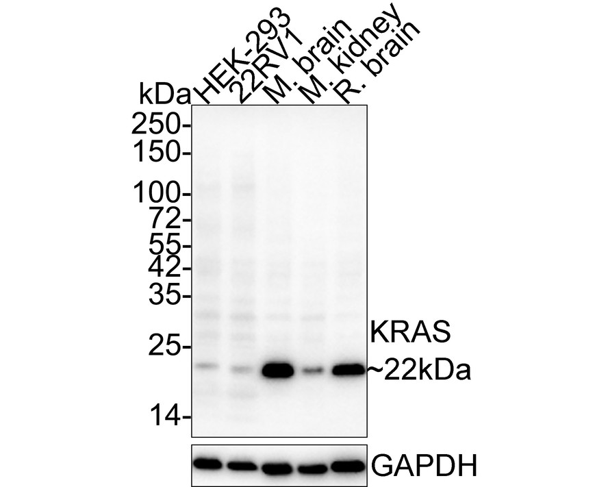
Western blot analysis of KRAS on different lysates with Mouse anti-KRAS antibody at 1/2,000 dilution. Lane 1: HEK-293 cell lysate, Lane 2: 22RV1 cell lysate, Lane 3: Mouse brain tissue lysate, Lane 4: Mouse kidney tissue lysate, Lane 5: Rat brain tissue lysate, Lysates/proteins at 30 µg/Lane. Exposure time: 43 seconds; 4-20% SDS-PAGE gel. Proteins were transferred to a PVDF membrane and blocked with 5% NFDM/TBST for 1 hour at room temperature. The primary antibody at 1/2,000 dilution was used in 5% NFDM/TBST at 4℃ overnight. Anti-Mouse IgG for IP Nano-secondary antibody at 1/5,000 dilution was used for 1 hour at room temperature.IHC
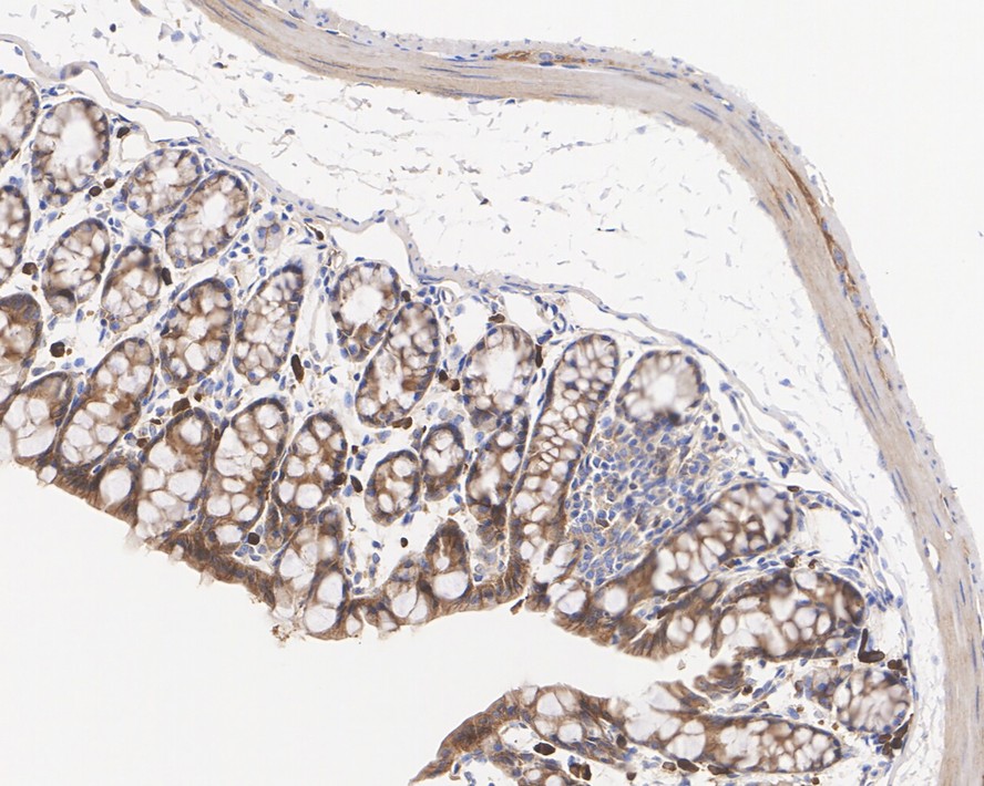
Immunohistochemical analysis of paraffin-embedded mouse colon tissue with Mouse anti-KRAS antibody at 1/1,000 dilution. The section was pre-treated using heat mediated antigen retrieval with Tris-EDTA buffer (pH 9.0) for 20 minutes. The tissues were blocked in 1% BSA for 20 minutes at room temperature, washed with ddH2O and PBS, and then probed with the primary antibody at 1/1,000 dilution for 1 hour at room temperature. The detection was performed using an HRP conjugated compact polymer system. DAB was used as the chromogen. Tissues were counterstained with hematoxylin and mounted with DPX.IHC
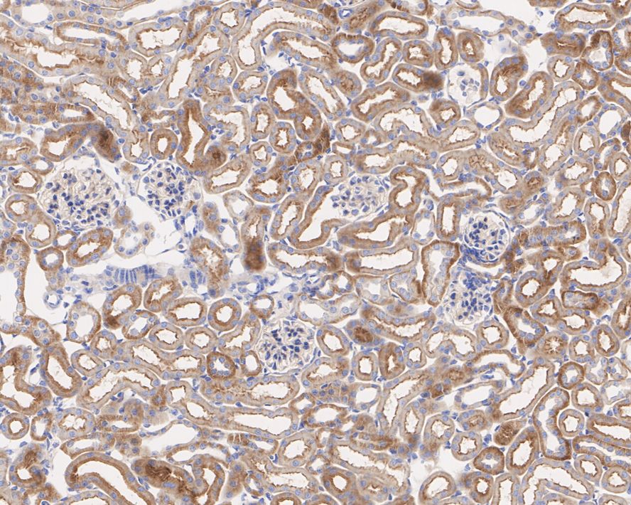
Immunohistochemical analysis of paraffin-embedded mouse kidney tissue with Mouse anti-KRAS antibody at 1/1,000 dilution. The section was pre-treated using heat mediated antigen retrieval with Tris-EDTA buffer (pH 9.0) for 20 minutes. The tissues were blocked in 1% BSA for 20 minutes at room temperature, washed with ddH2O and PBS, and then probed with the primary antibody at 1/1,000 dilution for 1 hour at room temperature. The detection was performed using an HRP conjugated compact polymer system. DAB was used as the chromogen. Tissues were counterstained with hematoxylin and mounted with DPX.IHC
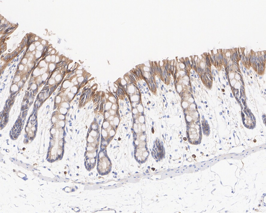
Immunohistochemical analysis of paraffin-embedded rat colon tissue with Mouse anti-KRAS antibody at 1/1,000 dilution. The section was pre-treated using heat mediated antigen retrieval with Tris-EDTA buffer (pH 9.0) for 20 minutes. The tissues were blocked in 1% BSA for 20 minutes at room temperature, washed with ddH2O and PBS, and then probed with the primary antibody at 1/1,000 dilution for 1 hour at room temperature. The detection was performed using an HRP conjugated compact polymer system. DAB was used as the chromogen. Tissues were counterstained with hematoxylin and mounted with DPX.| Product Name | KRAS Recombinant Mouse Monoclonal Antibody |
|---|---|
| Antibody Type | Primary Antibodies |
| Immunogen | Recombinant protein within human KRAS aa 2-186. |
| Clonality | Monoclonal |
|---|---|
| Isotype | IgG1 |
| Host Species | Mouse |
| Tested Applications | IHCWB |
| WB:1:500-1:2000 IHC:1:1000 |
|
| Species Reactivity | HumanMouseRat |
| Concentration | 1mg/ml |
| Purification | Protein A |
| Gene Symbol | KRAS |
|---|---|
| Gene Synonyms | NS NS3 OES CFC2 RALD K-Ras KRAS1 KRAS2 RALD2 RASK2 KI-RAS C-K-RAS K-RAS2A K-RAS2B K-RAS4A K-RAS4B K-Ras 2 'C-K-RAS c-Ki-ras c-Ki-ras2 |
| Gene Full Name | KRAS proto-oncogene, GTPase |
| Gene Summary | This gene, a Kirsten ras oncogene homolog from the mammalian ras gene family, encodes a protein that is a member of the small GTPase superfamily. A single amino acid substitution is responsible for an activating mutation. The transforming protein that results is implicated in various malignancies, including lung adenocarcinoma, mucinous adenoma, ductal carcinoma of the pancreas and colorectal carcinoma. Alternative splicing leads to variants encoding two isoforms that differ in the C-terminal region. [provided by RefSeq, Jul 2008] |
| Alternative Names | c Ki ras2 antibody c Kirsten ras protein antibody c-K-ras antibody c-Ki-ras antibody Cellular c Ki ras2 proto oncogene antibody Cellular transforming proto oncogene antibody CFC2 antibody cK Ras antibody GTPase KRas antibody K RAS p21 protein antibody c Ki ras2 antibody c Kirsten ras protein antibody c-K-ras antibody c-Ki-ras antibody Cellular c Ki ras2 proto oncogene antibody Cellular transforming proto oncogene antibody CFC2 antibody cK Ras antibody GTPase KRas antibody K RAS p21 protein antibody K RAS2A antibody K RAS2B antibody K RAS4A antibody K RAS4B antibody K-Ras 2 antibody KI RAS antibody Ki-Ras antibody KIRSTEN MURINE SARCOMA VIRUS 2 antibody Kirsten rat sarcoma 2 viral (v Ki ras2) oncogene homolog antibody Kirsten rat sarcoma viral oncogene homolog antibody KRAS antibody KRAS proto oncogene GTPase antibody KRAS1 antibody KRAS2 antibody N-terminally processed antibody NS antibody NS3 antibody Oncogene KRAS2 antibody p21ras antibody PR310 c K ras oncogene antibody PR310 cK ras oncogene antibody RALD antibody RASK_HUMAN antibody RASK2 antibody Transforming protein p21 antibody v Ki ras2 Kirsten rat sarcoma 2 viral oncogene homolog antibody v Ki ras2 Kirsten rat sarcoma viral oncogene homolog antibody |
| Molecular Weight(MW) | 22 kDa |
| Cellular Localization | Cell membrane. Cytoplasm. |
WB
Western blot analysis of KRAS on different lysates with Mouse anti-KRAS antibody at 1/2,000 dilution. Lane 1: HEK-293 cell lysate, Lane 2: 22RV1 cell lysate, Lane 3: Mouse brain tissue lysate, Lane 4: Mouse kidney tissue lysate, Lane 5: Rat brain tissue lysate, Lysates/proteins at 30 µg/Lane. Exposure time: 43 seconds; 4-20% SDS-PAGE gel. Proteins were transferred to a PVDF membrane and blocked with 5% NFDM/TBST for 1 hour at room temperature. The primary antibody at 1/2,000 dilution was used in 5% NFDM/TBST at 4℃ overnight. Anti-Mouse IgG for IP Nano-secondary antibody at 1/5,000 dilution was used for 1 hour at room temperature.
IHC
Immunohistochemical analysis of paraffin-embedded mouse colon tissue with Mouse anti-KRAS antibody at 1/1,000 dilution. The section was pre-treated using heat mediated antigen retrieval with Tris-EDTA buffer (pH 9.0) for 20 minutes. The tissues were blocked in 1% BSA for 20 minutes at room temperature, washed with ddH2O and PBS, and then probed with the primary antibody at 1/1,000 dilution for 1 hour at room temperature. The detection was performed using an HRP conjugated compact polymer system. DAB was used as the chromogen. Tissues were counterstained with hematoxylin and mounted with DPX.
IHC
Immunohistochemical analysis of paraffin-embedded mouse kidney tissue with Mouse anti-KRAS antibody at 1/1,000 dilution. The section was pre-treated using heat mediated antigen retrieval with Tris-EDTA buffer (pH 9.0) for 20 minutes. The tissues were blocked in 1% BSA for 20 minutes at room temperature, washed with ddH2O and PBS, and then probed with the primary antibody at 1/1,000 dilution for 1 hour at room temperature. The detection was performed using an HRP conjugated compact polymer system. DAB was used as the chromogen. Tissues were counterstained with hematoxylin and mounted with DPX.
IHC
Immunohistochemical analysis of paraffin-embedded rat colon tissue with Mouse anti-KRAS antibody at 1/1,000 dilution. The section was pre-treated using heat mediated antigen retrieval with Tris-EDTA buffer (pH 9.0) for 20 minutes. The tissues were blocked in 1% BSA for 20 minutes at room temperature, washed with ddH2O and PBS, and then probed with the primary antibody at 1/1,000 dilution for 1 hour at room temperature. The detection was performed using an HRP conjugated compact polymer system. DAB was used as the chromogen. Tissues were counterstained with hematoxylin and mounted with DPX.| Application Notes | WB:1:500-1:2000 IHC:1:1000 |
|---|
| Form | Liquid |
|---|---|
| Storage Instructions | Shipped at 4℃. Store at +4℃ short term (1-2 weeks). It is recommended to aliquot into single-use upon delivery. Store at -20℃ long term. |
| Storage Buffer | PBS (pH7.4), 0.1% BSA, 40% Glycerol. Preservative: 0.05% Sodium Azide.. |


