WB
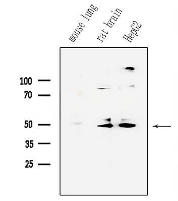
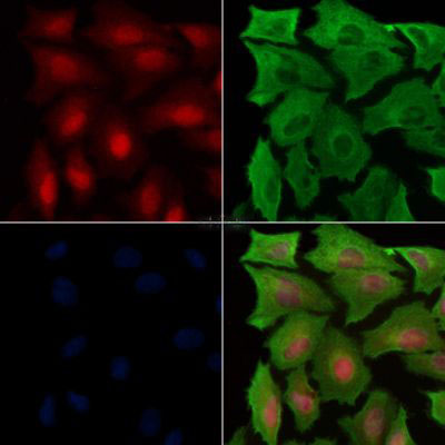
Western blot analysis of mouse brian lysates using Parkin Antibody. The lane on the left was treated with the antigen-specific peptide.Phospho-Parkin (Ser65) Antibody staining A549 cells(H2O2 treatment) by ICC/IF. The samples were fixed with PFA and permeabilized in 0.1% Triton X-100, then blocked in 10% serum for 45 minutes at 25°C. Samples were then incubated with primary Ab and mouse anti-beta tubulin Ab for 1 hour at 37°C. An AlexaFluor594 conjugated goat anti-rabbit IgG Ab(Red) and an AlexaFluor488 conjugated goat anti-mouse IgG Ab(Green) were used as the secondary antibody. The nuclear counter stain is DAPI (blue).
| Product Name | Rabbit polyclonal antibody to Phospho-Parkin (Ser65) |
|---|---|
| Antibody Type | Primary Antibodies |
| Immunogen | A synthesized peptide derived from human Parkin around the phosphorylation site of Ser65. |
| Modification | p-Ser65 |
| Clonality | Polyclonal |
|---|---|
| Isotype | IgG |
| Host Species | Rabbit |
| Tested Applications | ICC/IFWB |
| ICC/IF:1:100-1:500 WB:1:500-2000 |
|
| Species Reactivity | HumanMouseRat |
| Concentration | 1mg/ml |
| Purification | Affinity purified |
| Gene Symbol | PRKN |
|---|---|
| Gene Synonyms | PDJ AR-JP LPRS2 PARK2 |
| Gene Full Name | parkin RBR E3 ubiquitin protein ligase |
| Gene Summary | The precise function of this gene is unknown; however, the encoded protein is a component of a multiprotein E3 ubiquitin ligase complex that mediates the targeting of substrate proteins for proteasomal degradation. Mutations in this gene are known to cause Parkinson disease and autosomal recessive juvenile Parkinson disease. Alternative splicing of this gene produces multiple transcript variants encoding distinct isoforms. Additional splice variants of this gene have been described but currently lack transcript support. [provided by RefSeq, Jul 2008] |
| Alternative Names | AR JP E3 ubiquitin ligase E3 ubiquitin protein ligase parkin E3 ubiquitin-protein ligase parkin FRA6E LPRS 2 LPRS2 PARK 2 Park2 Parkin 2 Parkinson disease (autosomal recessive juvenile) 2 Parkinson disease (autosomal recessive juvenile) 2 parkin Parkinson disease protein 2 Parkinson juvenile disease protein 2 Parkinson protein 2 E3 ubiquitin protein ligase Parkinson protein 2 E3 ubiquitin protein ligase (parkin) PDJ PRKN 2 PRKN PRKN2 PRKN2_HUMAN Ubiquitin E3 ligase PRKN |
| Molecular Weight(MW) | 52 kDa |
| Cellular Localization | Cytoplasm>Cytosol. Nucleus. Endoplasmic reticulum. Mitochondrion. |
WB
Western blot analysis of mouse brian lysates using Parkin Antibody. The lane on the left was treated with the antigen-specific peptide.
Phospho-Parkin (Ser65) Antibody staining A549 cells(H2O2 treatment) by ICC/IF. The samples were fixed with PFA and permeabilized in 0.1% Triton X-100, then blocked in 10% serum for 45 minutes at 25°C. Samples were then incubated with primary Ab and mouse anti-beta tubulin Ab for 1 hour at 37°C. An AlexaFluor594 conjugated goat anti-rabbit IgG Ab(Red) and an AlexaFluor488 conjugated goat anti-mouse IgG Ab(Green) were used as the secondary antibody. The nuclear counter stain is DAPI (blue).
| Application Notes | ICC/IF:1:100-1:500 WB:1:500-2000 |
|---|
| Form | Liquid |
|---|---|
| Storage Instructions | Store at -20 °C. Stable for 12 months from date of receipt. |
| Storage Buffer | Rabbit IgG in phosphate buffered saline , pH 7.4, 150mM NaCl, 0.02% sodium azide and 50% glycerol. |


