WB
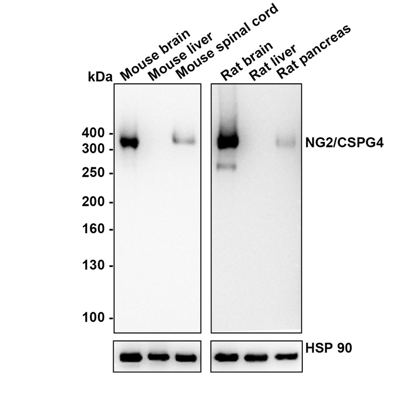
Western blot analysis of NG2/CSPG4 on different lysates with Rabbit anti-NG2/CSPG4 antibody at 1/5,000 dilution. Lysates/proteins at 30 µg/Lane. Exposure time: 30 seconds (mouse), 80 seconds (rat); Liver is a negative control. Blocking: 5% NFDM/TBST, 1 hour at room temperature. Primary antibody: Rabbit anti-NG2/CSPG4 antibody, 1/5,000 in primary antibody dilution buffer (K1803), overnight at 4 ℃. Secondary antibody: Goat anti-Rabbit IgG-HRP, 1/50,000 in 5% NFDM/TBST, 1 hour at room temperatureICC/IF
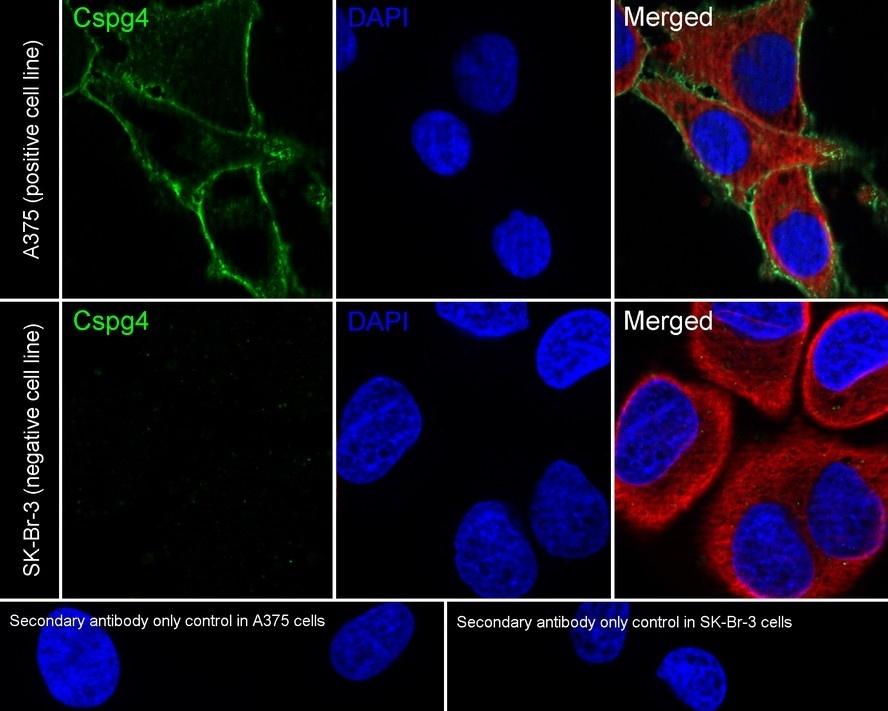
Application: Immunocytochemistry (IF-cell) Species: Human. Sample: A375 (Human amelanotic melanoma cells)/SK-BR-3 (Human breast cancer cells). Fixation: 4% Paraformaldehyde, 15 minutes at room temperature. Permeabilization: 0.1% Triton X-100, 15 minutes at room temperature. Blocking: 1% BSA + 10% normal goat serum, 1 hour at room temperature. Antibody dilution buffer: 1% BSA in PBST. Primary antibody: Rabbit anti-NG2/CSPG4 antibody, 1/200, overnight at 4°C. Secondary antibody: Goat Anti-Rabbit IgG (488), 45 minutes at room temperature. Counterstain: Beta tubulin (red), 1/100, overnight at 4℃. The Nuclear counterstain was DAPI (Blue). Negative control: SK-Br-3.IHC
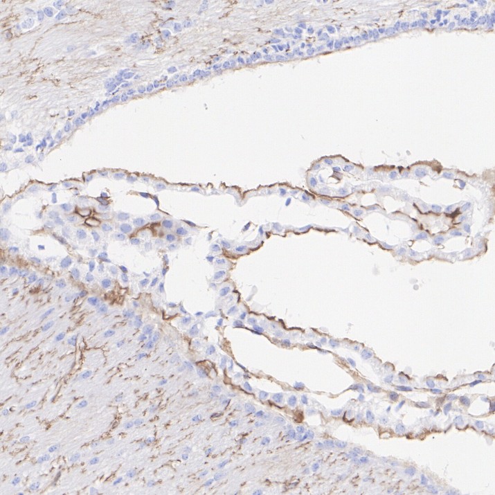
Application: Immunohistochemistry (IHC-P). Species: Mouse. Tissue: Choroid plexus. Sample: Paraffin-embedded section. Antigen retrieval: Heat-mediated, Tris-EDTA buffer (pH 9.0), 20 minutes at 95°C. Wash buffer: 1× TBST. Endogenous peroxidase blocking: 3% H₂O₂, 10 minutes at room temperature. Blocking: 1% BSA + 10% normal goat serum, 10 minutes at room temperature. Primary antibody: 1/1,000, 1 hour at room temperature. Secondary antibody: 20 minutes at room temperature.IHC
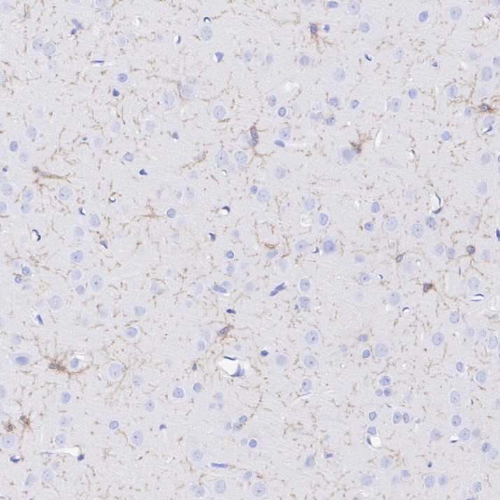
Application: Immunohistochemistry (IHC-P). Species: Rat. Tissue: Brain. Sample: Paraffin-embedded section. Antigen retrieval: Heat-mediated, Tris-EDTA buffer (pH 9.0), 20 minutes at 95°C. Wash buffer: 1× TBST. Endogenous peroxidase blocking: 3% H₂O₂, 10 minutes at room temperature. Blocking: 1% BSA + 10% normal goat serum, 10 minutes at room temperature. Primary antibody: 1/1,000, 1 hour at room temperature. Secondary antibody: 20 minutes at room temperature.IF-P
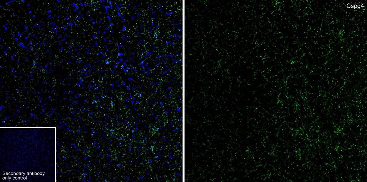
Application: Immunofluorescence (IF-tissue). Species: Mouse. Tissue: Brain. Sample: Paraffin-embedded section. Antigen retrieval: Heat-mediated, Tris-EDTA buffer (pH 9.0), 20 minutes at 95°C. Wash buffer: 1× TBST. Blocking: 10% normal goat serum + 1% Triton X-100 + 0.3 M Glycine in TBST, 30 minutes at room temperature. Primary antibody: 1/200, overnight at 4°C. Secondary antibody: Goat Anti-Rabbit IgG (488), 1.5 hours at room temperature.IF-P
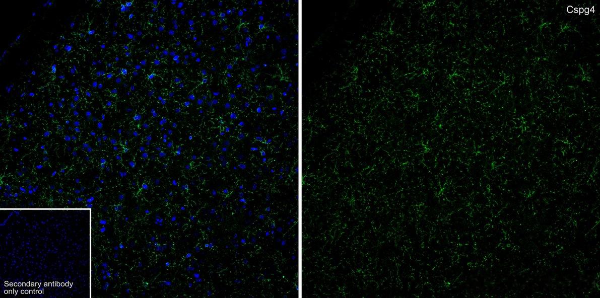
Application: Immunofluorescence (IF-tissue). Species: Rat. Tissue: Brain. Sample: Paraffin-embedded section. Antigen retrieval: Heat-mediated, Tris-EDTA buffer (pH 9.0), 20 minutes at 95°C. Wash buffer: 1× TBST Blocking: 10% normal goat serum + 1% Triton X-100 + 0.3 M Glycine in TBST, 30 minutes at room temperature. Primary antibody: 1/200, overnight at 4°C. Secondary antibody: Goat Anti-Rabbit IgG (488), 1.5 hours at room temperature.FC
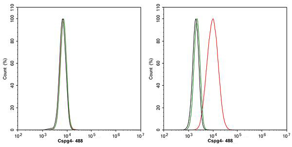
Application: Flow Cytometry. Species: Human . Sample: A375 (Human amelanotic melanoma cells)/SK-BR-3 (Human breast cancer cells). Blocking: 1% BSA + 10% normal goat serum, 15 minutes at room temperature. Antibody dilution buffer: 1x PBS. Primary antibody: Rabbit anti-NG2/CSPG4 antibody (1/1,000) (Red) compared with Rabbit IgG Isotype Control (Green), 15 minutes at room temperature. Secondary antibody: Goat Anti-Rabbit IgG (488), 15 minutes at room temperature. Negative control: SK-Br-3.| Product Name | NG2/CSPG4 Recombinant Rabbit Monoclonal Antibody |
|---|---|
| Antibody Type | Primary Antibodies |
| Immunogen | Recombinant protein within mouse CSPG4 aa 151-600. |
| Clonality | Monoclonal |
|---|---|
| Isotype | IgG |
| Host Species | Rabbit |
| Tested Applications | FCICC/IFIF-PIHCWB |
| WB:1:5000 ICC/IF:1:200 IHC:1:1000 IF-P:1:200 FC:1:1000 |
|
| Species Reactivity | HumanMouseRat |
| Concentration | 1mg/ml |
| Purification | Protein A |
| Gene Symbol | CSPG4 |
|---|---|
| Gene Synonyms | NG2 MCSP MCSPG MSK16 CSPG4A HMW-MAA MEL-CSPG |
| Gene Full Name | chondroitin sulfate proteoglycan 4 |
| Gene Summary | A human melanoma-associated chondroitin sulfate proteoglycan plays a role in stabilizing cell-substratum interactions during early events of melanoma cell spreading on endothelial basement membranes. CSPG4 represents an integral membrane chondroitin sulfate proteoglycan expressed by human malignant melanoma cells. [provided by RefSeq, Jul 2008] |
| Alternative Names | 4732461B14Rik antibody AN2 antibody AN2 proteoglycan antibody Chondroitin sulfate proteoglycan 4 (melanoma-associated) antibody Chondroitin sulfate proteoglycan 4 antibody Chondroitin sulfate proteoglycan NG2 antibody Cspg4 antibody Cspg4 chondroitin sulfate proteoglycan 4 antibody CSPG4_HUMAN antibody HMW-MAA antibody HSN tumor-specific antigen antibody Kiaa4232 antibody MCSP antibody MCSPG antibody MEL-CSPG antibody Melanoma chondroitin sulfate proteoglycan antibody Melanoma-associated chondroitin sulfate proteoglycan antibody MELCSPG antibody MSK16 antibody NG2 antibody |
| Molecular Weight(MW) | 251 kDa(Observed band size: 330 kDa) |
| Cellular Localization | Cell membrane, Apical cell membrane, Cell projection, lamellipodium membrane, Cell surface. |
WB
Western blot analysis of NG2/CSPG4 on different lysates with Rabbit anti-NG2/CSPG4 antibody at 1/5,000 dilution. Lysates/proteins at 30 µg/Lane. Exposure time: 30 seconds (mouse), 80 seconds (rat); Liver is a negative control. Blocking: 5% NFDM/TBST, 1 hour at room temperature. Primary antibody: Rabbit anti-NG2/CSPG4 antibody, 1/5,000 in primary antibody dilution buffer (K1803), overnight at 4 ℃. Secondary antibody: Goat anti-Rabbit IgG-HRP, 1/50,000 in 5% NFDM/TBST, 1 hour at room temperature
ICC/IF
Application: Immunocytochemistry (IF-cell) Species: Human. Sample: A375 (Human amelanotic melanoma cells)/SK-BR-3 (Human breast cancer cells). Fixation: 4% Paraformaldehyde, 15 minutes at room temperature. Permeabilization: 0.1% Triton X-100, 15 minutes at room temperature. Blocking: 1% BSA + 10% normal goat serum, 1 hour at room temperature. Antibody dilution buffer: 1% BSA in PBST. Primary antibody: Rabbit anti-NG2/CSPG4 antibody, 1/200, overnight at 4°C. Secondary antibody: Goat Anti-Rabbit IgG (488), 45 minutes at room temperature. Counterstain: Beta tubulin (red), 1/100, overnight at 4℃. The Nuclear counterstain was DAPI (Blue). Negative control: SK-Br-3.
IHC
Application: Immunohistochemistry (IHC-P). Species: Mouse. Tissue: Choroid plexus. Sample: Paraffin-embedded section. Antigen retrieval: Heat-mediated, Tris-EDTA buffer (pH 9.0), 20 minutes at 95°C. Wash buffer: 1× TBST. Endogenous peroxidase blocking: 3% H₂O₂, 10 minutes at room temperature. Blocking: 1% BSA + 10% normal goat serum, 10 minutes at room temperature. Primary antibody: 1/1,000, 1 hour at room temperature. Secondary antibody: 20 minutes at room temperature.
IHC
Application: Immunohistochemistry (IHC-P). Species: Rat. Tissue: Brain. Sample: Paraffin-embedded section. Antigen retrieval: Heat-mediated, Tris-EDTA buffer (pH 9.0), 20 minutes at 95°C. Wash buffer: 1× TBST. Endogenous peroxidase blocking: 3% H₂O₂, 10 minutes at room temperature. Blocking: 1% BSA + 10% normal goat serum, 10 minutes at room temperature. Primary antibody: 1/1,000, 1 hour at room temperature. Secondary antibody: 20 minutes at room temperature.
IF-P
Application: Immunofluorescence (IF-tissue). Species: Mouse. Tissue: Brain. Sample: Paraffin-embedded section. Antigen retrieval: Heat-mediated, Tris-EDTA buffer (pH 9.0), 20 minutes at 95°C. Wash buffer: 1× TBST. Blocking: 10% normal goat serum + 1% Triton X-100 + 0.3 M Glycine in TBST, 30 minutes at room temperature. Primary antibody: 1/200, overnight at 4°C. Secondary antibody: Goat Anti-Rabbit IgG (488), 1.5 hours at room temperature.
IF-P
Application: Immunofluorescence (IF-tissue). Species: Rat. Tissue: Brain. Sample: Paraffin-embedded section. Antigen retrieval: Heat-mediated, Tris-EDTA buffer (pH 9.0), 20 minutes at 95°C. Wash buffer: 1× TBST Blocking: 10% normal goat serum + 1% Triton X-100 + 0.3 M Glycine in TBST, 30 minutes at room temperature. Primary antibody: 1/200, overnight at 4°C. Secondary antibody: Goat Anti-Rabbit IgG (488), 1.5 hours at room temperature.
FC
Application: Flow Cytometry. Species: Human . Sample: A375 (Human amelanotic melanoma cells)/SK-BR-3 (Human breast cancer cells). Blocking: 1% BSA + 10% normal goat serum, 15 minutes at room temperature. Antibody dilution buffer: 1x PBS. Primary antibody: Rabbit anti-NG2/CSPG4 antibody (1/1,000) (Red) compared with Rabbit IgG Isotype Control (Green), 15 minutes at room temperature. Secondary antibody: Goat Anti-Rabbit IgG (488), 15 minutes at room temperature. Negative control: SK-Br-3.| Application Notes | WB:1:5000 ICC/IF:1:200 IHC:1:1000 IF-P:1:200 FC:1:1000 |
|---|
| Form | Liquid |
|---|---|
| Storage Instructions | Shipped at 4℃. Store at +4℃ short term (1-2 weeks). It is recommended to aliquot into single-use upon delivery. Store at -20℃ long term. |
| Storage Buffer | PBS (pH7.4), 0.1% BSA, 40% Glycerol. Preservative: 0.05% Sodium Azide. |


