WB
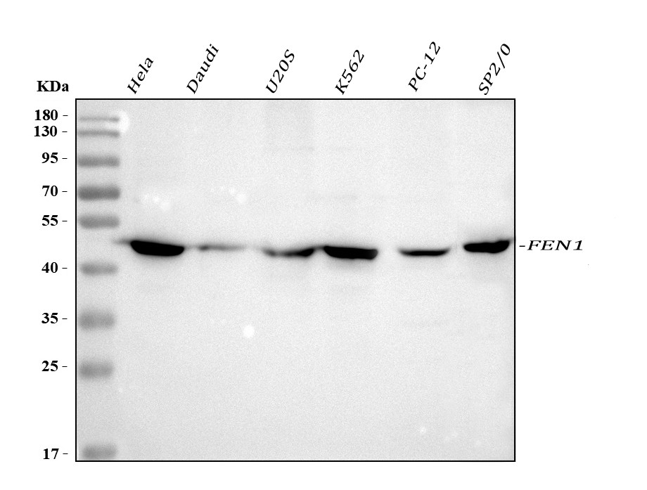
Western blot analysis of FEN1 using anti-FEN1 antibody. The sample well of each lane was loaded with 30 ug of sample under reducing conditions. After electrophoresis, proteins were transferred to a membrane. Then the membrane was incubated with rabbit anti-FEN1 antigen affinity purified polyclonal antibody at a dilution of 1:1000 and probed with a goat anti-rabbit IgG-HRP secondary antibody. The signal is developed using ECL Plus Western Blotting Substrate.IHC
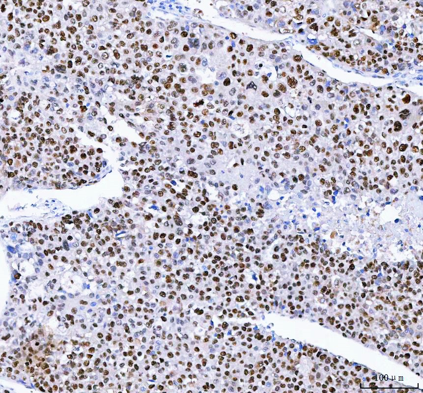
IHC analysis of FEN1 using anti-FEN1 antibody. FEN1 was detected in a paraffin-embedded section of human liver cancer tissue. The tissue section was incubated with rabbit anti-FEN1 Antibody at a dilution of 1:200 and developed using HRP Conjugated Rabbit IgG Super Vision Assay Kit with DAB as the chromogen.ICC/IF
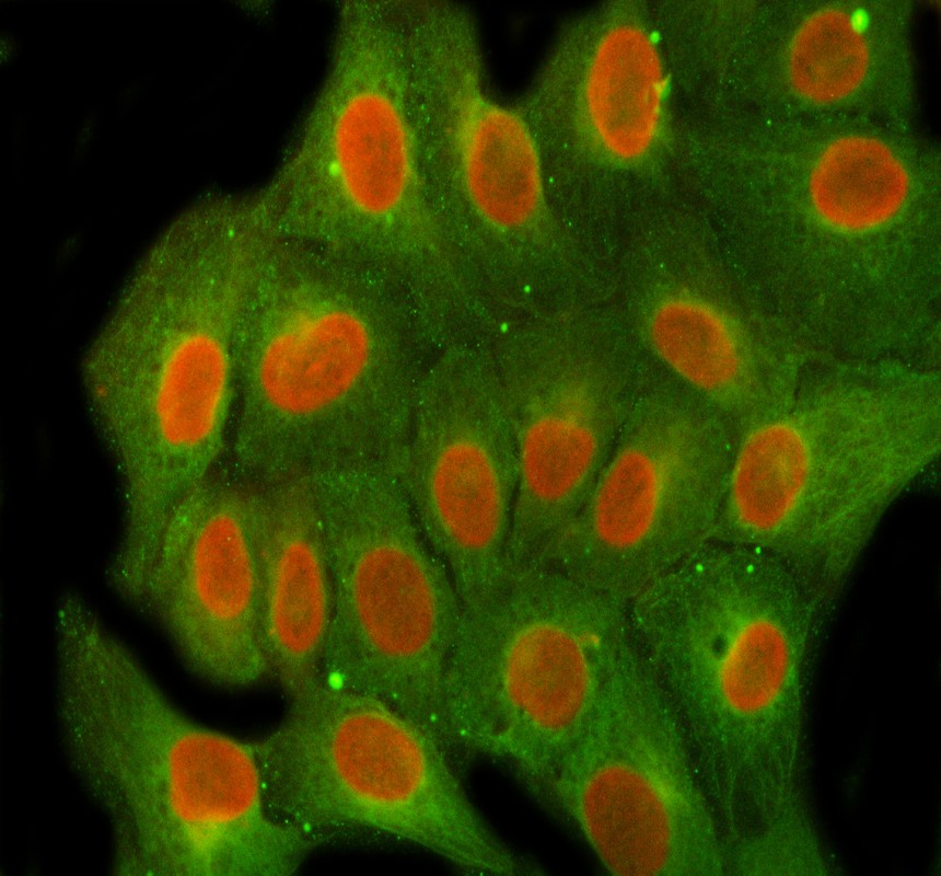
ICC/IF analysis of FEN1 using anti-FEN1 antibody and anti-Beta Tubulin antibody. FEN1 was detected in an immunocytochemical section of U2OS cells. The section was incubated with rabbit anti-FEN1 Antibody at a dilution of 1:100. Cy3-Conjugated Anti-rabbit IgG Secondary Antibody (Red) and Fluoro488-conjugated Anti-mouse IgG Secondary Antibody (Green) were used as secondary antibody.IF-P
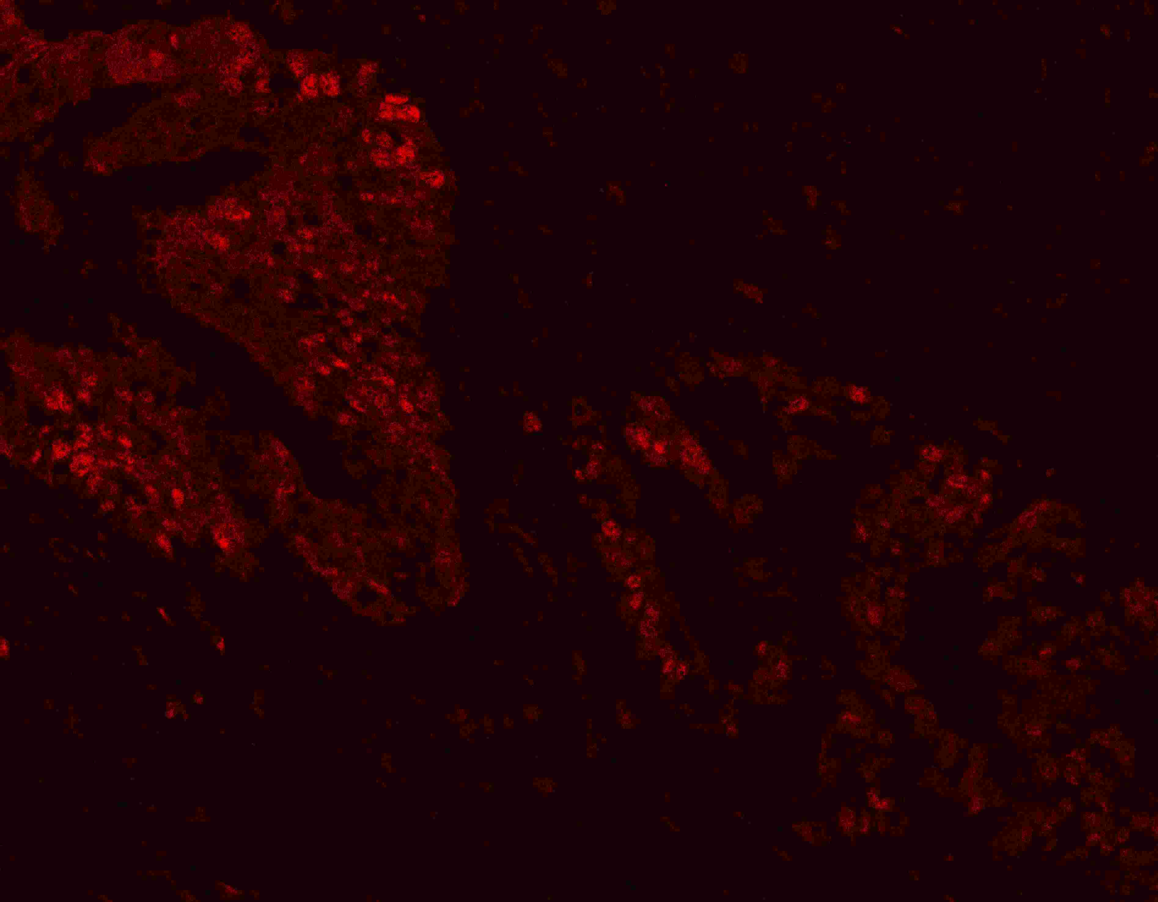
IF analysis of FEN1 using anti-FEN1 antibody. FEN1 was detected in a paraffin-embedded section of human intestinal cancer tissue. The tissue section was incubated with rabbit anti-FEN1 Antibody at a dilution of 1:100. Cy3-conjugated Anti-rabbit IgG Secondary Antibody (red) was used as secondary antibody.FC
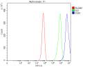
Flow Cytometry analysis of K562 cells using anti-FEN1 antibody. Overlay histogram showing K562 cells stained with Rabbit polyclonal antibody to FEN1 (Blue line). To facilitate intracellular staining, cells were fixed with 4% paraformaldehyde and permeabilized with permeabilization buffer. The cells were blocked with 10% normal goat serum. And then incubated with rabbit anti-FEN1 Antibody Rabbit polyclonal antibody to FEN1 at 1:100 dilution for 30 min at 20°C. Fluoro488 conjugated goat anti-rabbit IgG was used as secondary antibody at 1:100 dilution for 30 minutes at 20°C. Isotype control antibody (Green line) was rabbit IgG (1 μg/1x106) used under the same conditions. Unlabelled sample (Red line) was also used as a control.| Product Name | Rabbit polyclonal antibody to FEN1 |
|---|---|
| Antibody Type | Primary Antibodies |
| Immunogen | E.coli-derived human FEN1 recombinant protein (Position: Q4-E300). |
| Clonality | Polyclonal |
|---|---|
| Isotype | IgG |
| Host Species | Rabbit |
| Tested Applications | FCICC/IFIF-PIHCWB |
| WB:1:1000-1:5000 IHC:1:50-1:400 ICC/IF:1:50-1:400 IF-P:1:50-1:400 FC:1:50-1:200 |
|
| Species Reactivity | HumanMouseRat |
| Concentration | 0.5mg/ml |
| Purification | Affinity purified |
| Gene Symbol | FEN1 |
|---|---|
| Gene Synonyms | MF1 RAD2 FEN-1 |
| Gene Full Name | flap structure-specific endonuclease 1 |
| Gene Summary | The protein encoded by this gene removes 5' overhanging flaps in DNA repair and processes the 5' ends of Okazaki fragments in lagging strand DNA synthesis. Direct physical interaction between this protein and AP endonuclease 1 during long-patch base excision repair provides coordinated loading of the proteins onto the substrate, thus passing the substrate from one enzyme to another. The protein is a member of the XPG/RAD2 endonuclease family and is one of ten proteins essential for cell-free DNA replication. DNA secondary structure can inhibit flap processing at certain trinucleotide repeats in a length-dependent manner by concealing the 5' end of the flap that is necessary for both binding and cleavage by the protein encoded by this gene. Therefore, secondary structure can deter the protective function of this protein, leading to site-specific trinucleotide expansions. [provided by RefSeq, Jul 2008] |
| Alternative Names | DNase IV FEN 1 FEN1 Flap endonuclease 1 flap structure endonuclease 1 hFEN 1 Maturation factor 1 MF1 RAD2 |
| Molecular Weight(MW) | 43 kDa(Observed band size:48 kDa) |
| Cellular Localization | Mitochondrion,Nucleus. |
WB
Western blot analysis of FEN1 using anti-FEN1 antibody. The sample well of each lane was loaded with 30 ug of sample under reducing conditions. After electrophoresis, proteins were transferred to a membrane. Then the membrane was incubated with rabbit anti-FEN1 antigen affinity purified polyclonal antibody at a dilution of 1:1000 and probed with a goat anti-rabbit IgG-HRP secondary antibody. The signal is developed using ECL Plus Western Blotting Substrate.
IHC
IHC analysis of FEN1 using anti-FEN1 antibody. FEN1 was detected in a paraffin-embedded section of human liver cancer tissue. The tissue section was incubated with rabbit anti-FEN1 Antibody at a dilution of 1:200 and developed using HRP Conjugated Rabbit IgG Super Vision Assay Kit with DAB as the chromogen.
ICC/IF
ICC/IF analysis of FEN1 using anti-FEN1 antibody and anti-Beta Tubulin antibody. FEN1 was detected in an immunocytochemical section of U2OS cells. The section was incubated with rabbit anti-FEN1 Antibody at a dilution of 1:100. Cy3-Conjugated Anti-rabbit IgG Secondary Antibody (Red) and Fluoro488-conjugated Anti-mouse IgG Secondary Antibody (Green) were used as secondary antibody.
IF-P
IF analysis of FEN1 using anti-FEN1 antibody. FEN1 was detected in a paraffin-embedded section of human intestinal cancer tissue. The tissue section was incubated with rabbit anti-FEN1 Antibody at a dilution of 1:100. Cy3-conjugated Anti-rabbit IgG Secondary Antibody (red) was used as secondary antibody.
FC
Flow Cytometry analysis of K562 cells using anti-FEN1 antibody. Overlay histogram showing K562 cells stained with Rabbit polyclonal antibody to FEN1 (Blue line). To facilitate intracellular staining, cells were fixed with 4% paraformaldehyde and permeabilized with permeabilization buffer. The cells were blocked with 10% normal goat serum. And then incubated with rabbit anti-FEN1 Antibody Rabbit polyclonal antibody to FEN1 at 1:100 dilution for 30 min at 20°C. Fluoro488 conjugated goat anti-rabbit IgG was used as secondary antibody at 1:100 dilution for 30 minutes at 20°C. Isotype control antibody (Green line) was rabbit IgG (1 μg/1x106) used under the same conditions. Unlabelled sample (Red line) was also used as a control.| Application Notes | WB:1:1000-1:5000 IHC:1:50-1:400 ICC/IF:1:50-1:400 IF-P:1:50-1:400 FC:1:50-1:200 |
|---|
| Form | Liquid |
|---|---|
| Storage Instructions | 12 months from date of receipt,-20℃ as supplied. |
| Storage Buffer | 500 ug/ml antibody with PBS, 0.02% NaN3, 1 mg/ml BSA and 50% glycerol. |


