WB
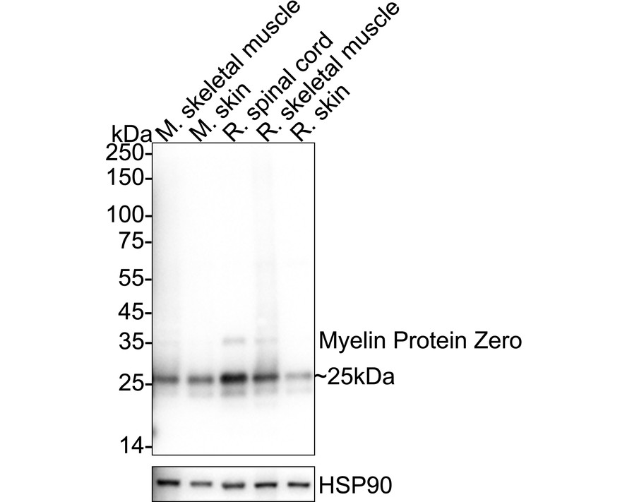
Western blot analysis of Myelin Protein Zero on different lysates with Rabbit anti-Myelin Protein Zero antibody at 1/2,000 dilution. Exposure time: 2 seconds; 4-20% SDS-PAGE gel. Proteins were transferred to a PVDF membrane and blocked with 5% NFDM/TBST for 1 hour at room temperature. The primary antibody at 1/2,000 dilution was used in primary antibody dilution at 4℃ overnight. Goat Anti-Rabbit IgG - HRP Secondary Antibody at 1/50,000 dilution was used for 1 hour at room temperature.IHC
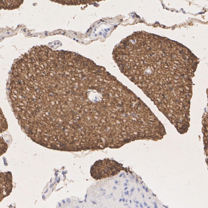
Immunohistochemical analysis of paraffin-embedded mouse dorsal root ganglion tissue with Rabbit anti-Myelin Protein Zero antibody at 1/1,000 dilution. The section was pre-treated using heat mediated antigen retrieval with Tris-EDTA buffer (pH 9.0) for 20 minutes. The tissues were blocked in 1% BSA for 20 minutes at room temperature, washed with ddH2O and PBS, and then probed with the primary antibody at 1/1,000 dilution for 1 hour at room temperature. The detection was performed using an HRP conjugated compact polymer system. DAB was used as the chromogen. Tissues were counterstained with hematoxylin and mounted with DPX.IHC
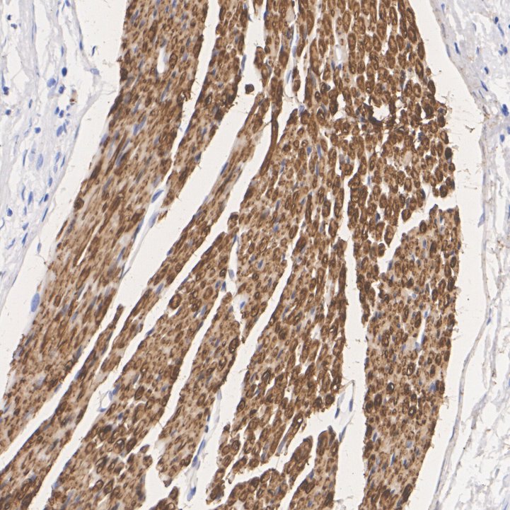
Immunohistochemical analysis of paraffin-embedded rat dorsal root ganglion tissue with Rabbit anti-Myelin Protein Zero antibody at 1/1,000 dilution. The section was pre-treated using heat mediated antigen retrieval with Tris-EDTA buffer (pH 9.0) for 20 minutes. The tissues were blocked in 1% BSA for 20 minutes at room temperature, washed with ddH2O and PBS, and then probed with the primary antibody at 1/1,000 dilution for 1 hour at room temperature. The detection was performed using an HRP conjugated compact polymer system. DAB was used as the chromogen. Tissues were counterstained with hematoxylin and mounted with DPX.IF-F
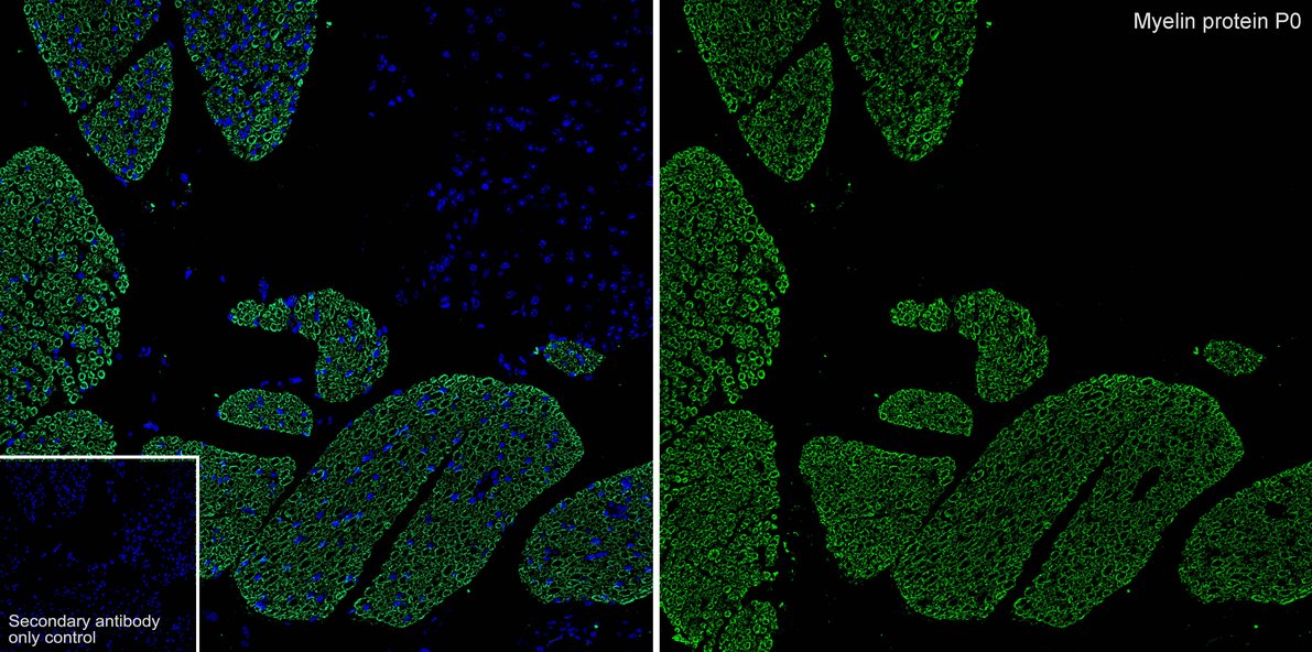
Application: IHC-Fr. Species: Mouse. Site: spinal cord. Sample: Frozen section. Antibody concentration: 1/500. Antigen retrieval: Not required.IF-P
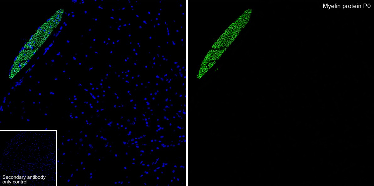
Application: IF-Tissue. Species: Mouse. Site: dorsal root ganglion. Sample: Paraffin-embedded section. Antibody concentration: 1/500.IP
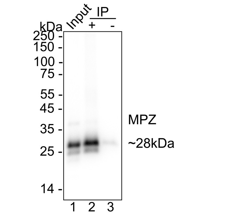
Myelin Protein Zero was immunoprecipitated from 0.2 mg Mouse spinal cord tissue lysate with Myelin Protein Zero Recombinant Rabbit Monoclonal Antibody at 2 µg/10 µl beads. Western blot was performed from the immunoprecipitate using Myelin Protein Zero Recombinant Rabbit Monoclonal Antibody at 1/1,000 dilution. HRP Conjugated Anti-Rabbit IgG for IP Nano-secondary antibody at 1/5,000 dilution was used for 1 hour at room temperature. Lane 1: Mouse spinal cord tissue lysate (input). Lane 2: Myelin Protein Zero Recombinant Rabbit Monoclonal Antibody IP in Mouse spinal cord tissue lysate. Lane 3: Rabbit IgG instead of Myelin Protein Zero Recombinant Rabbit Monoclonal Antibody in Mouse spinal cord tissue lysate. Blocking/Dilution buffer: primary antibody dilution. Exposure time: 5 seconds.| Product Name | Myelin Protein Zero Recombinant Rabbit Monoclonal Antibody |
|---|---|
| Antibody Type | Primary Antibodies |
| Clonality | Monoclonal |
|---|---|
| Isotype | IgG |
| Host Species | Rabbit |
| Tested Applications | IF-FIF-PIHCIPWB |
| WB:1:2000 IHC:1:1000 IF-P:1:500 IF-F:1:500 IP:1-2μg/sample |
|
| Species Reactivity | MouseRat |
| Concentration | 1mg/ml |
| Purification | Protein A |
| Gene Symbol | MPZ |
|---|---|
| Gene Synonyms | P0 DSS MPP CMT1 CMT1B CMT2I CMT2J CMT4E CMTDI3 CMTDID HMSNIB |
| Gene Full Name | myelin protein zero |
| Gene Summary | This gene is specifically expressed in Schwann cells of the peripheral nervous system and encodes a type I transmembrane glycoprotein that is a major structural protein of the peripheral myelin sheath. The encoded protein contains a large hydrophobic extracellular domain and a smaller basic intracellular domain, which are essential for the formation and stabilization of the multilamellar structure of the compact myelin. Mutations in this gene are associated with autosomal dominant form of Charcot-Marie-Tooth disease type 1 (CMT1B) and other polyneuropathies, such as Dejerine-Sottas syndrome (DSS) and congenital hypomyelinating neuropathy (CHN). A recent study showed that two isoforms are produced from the same mRNA by use of alternative in-frame translation termination codons via a stop codon readthrough mechanism. [provided by RefSeq, Oct 2015] |
| Alternative Names | Charcot Marie Tooth neuropathy 1B antibody CHM antibody CMT1 antibody CMT1B antibody CMT2I antibody CMT2J antibody CMT4E antibody CMTDI3 antibody CMTDID antibody DSS antibody HMSNIB antibody MPP antibody MPZ antibody Myelin peripheral protein antibody Myelin protein P0 antibody Myelin protein zero antibody MYP0_HUMAN antibody P0 antibody |
| Molecular Weight(MW) | 28 kDa(Observed band size:25 kDa) |
| Cellular Localization | Cell membrane. |
WB
Western blot analysis of Myelin Protein Zero on different lysates with Rabbit anti-Myelin Protein Zero antibody at 1/2,000 dilution. Exposure time: 2 seconds; 4-20% SDS-PAGE gel. Proteins were transferred to a PVDF membrane and blocked with 5% NFDM/TBST for 1 hour at room temperature. The primary antibody at 1/2,000 dilution was used in primary antibody dilution at 4℃ overnight. Goat Anti-Rabbit IgG - HRP Secondary Antibody at 1/50,000 dilution was used for 1 hour at room temperature.
IHC
Immunohistochemical analysis of paraffin-embedded mouse dorsal root ganglion tissue with Rabbit anti-Myelin Protein Zero antibody at 1/1,000 dilution. The section was pre-treated using heat mediated antigen retrieval with Tris-EDTA buffer (pH 9.0) for 20 minutes. The tissues were blocked in 1% BSA for 20 minutes at room temperature, washed with ddH2O and PBS, and then probed with the primary antibody at 1/1,000 dilution for 1 hour at room temperature. The detection was performed using an HRP conjugated compact polymer system. DAB was used as the chromogen. Tissues were counterstained with hematoxylin and mounted with DPX.
IHC
Immunohistochemical analysis of paraffin-embedded rat dorsal root ganglion tissue with Rabbit anti-Myelin Protein Zero antibody at 1/1,000 dilution. The section was pre-treated using heat mediated antigen retrieval with Tris-EDTA buffer (pH 9.0) for 20 minutes. The tissues were blocked in 1% BSA for 20 minutes at room temperature, washed with ddH2O and PBS, and then probed with the primary antibody at 1/1,000 dilution for 1 hour at room temperature. The detection was performed using an HRP conjugated compact polymer system. DAB was used as the chromogen. Tissues were counterstained with hematoxylin and mounted with DPX.
IF-F
Application: IHC-Fr. Species: Mouse. Site: spinal cord. Sample: Frozen section. Antibody concentration: 1/500. Antigen retrieval: Not required.
IF-P
Application: IF-Tissue. Species: Mouse. Site: dorsal root ganglion. Sample: Paraffin-embedded section. Antibody concentration: 1/500.
IP
Myelin Protein Zero was immunoprecipitated from 0.2 mg Mouse spinal cord tissue lysate with Myelin Protein Zero Recombinant Rabbit Monoclonal Antibody at 2 µg/10 µl beads. Western blot was performed from the immunoprecipitate using Myelin Protein Zero Recombinant Rabbit Monoclonal Antibody at 1/1,000 dilution. HRP Conjugated Anti-Rabbit IgG for IP Nano-secondary antibody at 1/5,000 dilution was used for 1 hour at room temperature. Lane 1: Mouse spinal cord tissue lysate (input). Lane 2: Myelin Protein Zero Recombinant Rabbit Monoclonal Antibody IP in Mouse spinal cord tissue lysate. Lane 3: Rabbit IgG instead of Myelin Protein Zero Recombinant Rabbit Monoclonal Antibody in Mouse spinal cord tissue lysate. Blocking/Dilution buffer: primary antibody dilution. Exposure time: 5 seconds.| Application Notes | WB:1:2000 IHC:1:1000 IF-P:1:500 IF-F:1:500 IP:1-2μg/sample |
|---|
| Form | Liquid |
|---|---|
| Storage Instructions | Shipped at 4℃. Store at +4℃ short term (1-2 weeks). Store at -20℃ long term. |
| Storage Buffer | 1*PBS (pH7.4), 0.1% BSA, 40% Glycerol, 0.2% Proclean 950. |


