WB
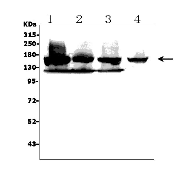
Western blot analysis of JMJD3/KDM6B using anti-JMJD3/KDM6B antibody. The sample well of each lane was loaded with 30 ug of sample under reducing conditions. Lane 1: human K562 whole cell lysates, Lane 2: human A375 whole cell lysates, Lane 3: human HEK293 whole cell lysates, Lane 4: human A431 whole cell lysates. After electrophoresis, proteins were transferred to a membrane. Then the membrane was incubated with rabbit anti-JMJD3/KDM6B antigen affinity purified polyclonal antibody at a dilution of 1:1000 and probed with a goat anti-rabbit IgG-HRP secondary antibody. The signal is developed using ECL Plus Western Blotting Substrate.FC
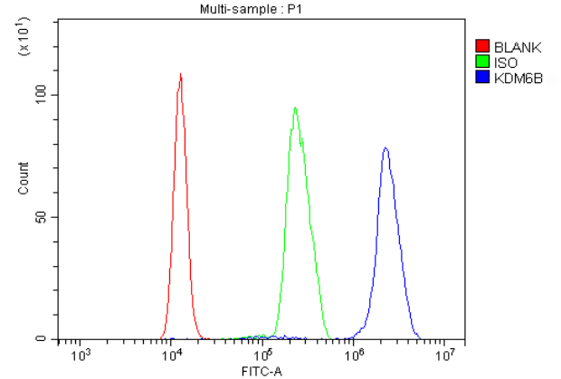
Flow Cytometry analysis of K562 cells using anti-JMJD3/KDM6B antibody. Overlay histogram showing K562 cells stained with Anti-JMJD3/KDM6B Antibody (Blue line). To facilitate intracellular staining, cells were fixed with 4% paraformaldehyde and permeabilized with permeabilization buffer. The cells were blocked with 10% normal goat serum. And then incubated with rabbit anti-JMJD3/KDM6B Antibody at 1:100 dilution for 30 min at 20°C. Fluoro488 conjugated goat anti-rabbit IgG was used as secondary antibody at 1:100 dilution for 30 minutes at 20°C. Isotype control antibody (Green line) was rabbit IgG at 1:100 dilution used under the same conditions. Unlabelled sample without incubation with primary antibody and secondary antibody (Red line) was used as a blank control.| Product Name | Rabbit polyclonal antibody to JMJD3/KDM6B |
|---|---|
| Antibody Type | Primary Antibodies |
| Immunogen | E.coli-derived human KDM6B/JMJD3 recombinant protein (Position: R1127-R1643). |
| Clonality | Polyclonal |
|---|---|
| Isotype | IgG |
| Host Species | Rabbit |
| Tested Applications | FCWB |
| WB:1:500-1:2000 FC:1:50-1:200 |
|
| Species Reactivity | HumanMouse |
| Concentration | 0.5mg/ml |
| Purification | Affinity purified |
| Gene Symbol | KDM6B |
|---|---|
| Gene Synonyms | JMJD3 NEDSST NEDCFSA |
| Gene Full Name | lysine demethylase 6B |
| Gene Summary | The protein encoded by this gene is a lysine-specific demethylase that specifically demethylates di- or tri-methylated lysine 27 of histone H3 (H3K27me2 or H3K27me3). H3K27 trimethylation is a repressive epigenetic mark controlling chromatin organization and gene silencing. This protein can also demethylate non-histone proteins such as retinoblastoma protein. Through its demethylation actvity this gene influences cellular differentiation and development, tumorigenesis, inflammatory diseases, and neurodegenerative diseases. This protein has two classical nuclear localization signals at its N-terminus. Alternative splicing results in multiple transcript variants encoding distinct isoforms. [provided by RefSeq, Feb 2017] |
| Alternative Names | lysine demethylase 6B JMJD3 NEDCFSA KDM6B KIAA0346 |
| Molecular Weight(MW) | 177 kDa |
| Cellular Localization | Nucleus. |
WB
Western blot analysis of JMJD3/KDM6B using anti-JMJD3/KDM6B antibody. The sample well of each lane was loaded with 30 ug of sample under reducing conditions. Lane 1: human K562 whole cell lysates, Lane 2: human A375 whole cell lysates, Lane 3: human HEK293 whole cell lysates, Lane 4: human A431 whole cell lysates. After electrophoresis, proteins were transferred to a membrane. Then the membrane was incubated with rabbit anti-JMJD3/KDM6B antigen affinity purified polyclonal antibody at a dilution of 1:1000 and probed with a goat anti-rabbit IgG-HRP secondary antibody. The signal is developed using ECL Plus Western Blotting Substrate.
FC
Flow Cytometry analysis of K562 cells using anti-JMJD3/KDM6B antibody. Overlay histogram showing K562 cells stained with Anti-JMJD3/KDM6B Antibody (Blue line). To facilitate intracellular staining, cells were fixed with 4% paraformaldehyde and permeabilized with permeabilization buffer. The cells were blocked with 10% normal goat serum. And then incubated with rabbit anti-JMJD3/KDM6B Antibody at 1:100 dilution for 30 min at 20°C. Fluoro488 conjugated goat anti-rabbit IgG was used as secondary antibody at 1:100 dilution for 30 minutes at 20°C. Isotype control antibody (Green line) was rabbit IgG at 1:100 dilution used under the same conditions. Unlabelled sample without incubation with primary antibody and secondary antibody (Red line) was used as a blank control.| Application Notes | WB:1:500-1:2000 FC:1:50-1:200 |
|---|
| Form | Liquid |
|---|---|
| Storage Instructions | 12 months from date of receipt,-20℃ as supplied. |
| Storage Buffer | 500 ug/ml antibody with PBS, 0.02% NaN3, 1 mg/ml BSA and 50% glycerol. |


