IHC
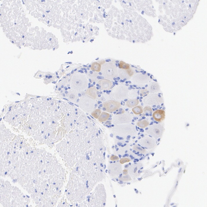
Immunohistochemical analysis of paraffin-embedded mouse dorsal root ganglion tissue with Rabbit anti-TRPV1 antibody at 1/15,000 dilution. The section was pre-treated using heat mediated antigen retrieval with Tris-EDTA buffer (pH 9.0) for 20 minutes. The tissues were blocked in 1% BSA for 20 minutes at room temperature, washed with ddH2O and PBS, and then probed with the primary antibody at 1/15,000 dilution for 1 hour at room temperature. The detection was performed using an HRP conjugated compact polymer system. DAB was used as the chromogen. Tissues were counterstained with hematoxylin and mounted with DPX.IHC
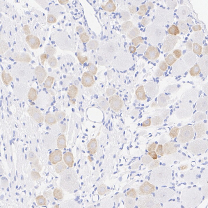
Immunohistochemical analysis of paraffin-embedded rat dorsal root ganglion tissue with Rabbit anti-TRPV1 antibody at 1/15,000 dilution. The section was pre-treated using heat mediated antigen retrieval with Tris-EDTA buffer (pH 9.0) for 20 minutes. The tissues were blocked in 1% BSA for 20 minutes at room temperature, washed with ddH2O and PBS, and then probed with the primary antibody at 1/15,000 dilution for 1 hour at room temperature. The detection was performed using an HRP conjugated compact polymer system. DAB was used as the chromogen. Tissues were counterstained with hematoxylin and mounted with DPX.IF-F
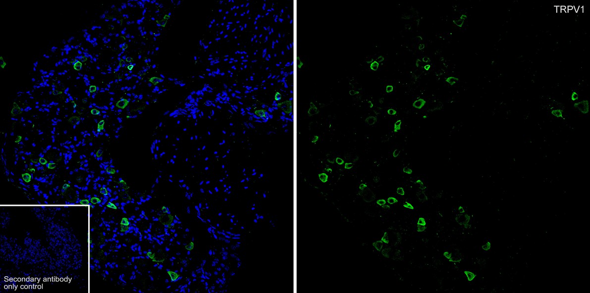
Application: IHC-Fr. Species: Mouse. Site: dorsal root ganglion. Sample: Frozen section. Antibody concentration: 1/1,000. Antigen retrieval: Not required.IF-F
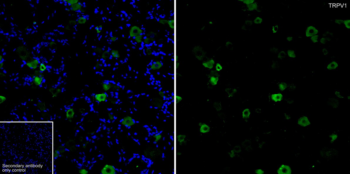
Application: IHC-Fr. Species: Rat. Site: dorsal root ganglion. Sample: Frozen section. Antibody concentration: 1/5,000. Antigen retrieval: Not required.IF-P
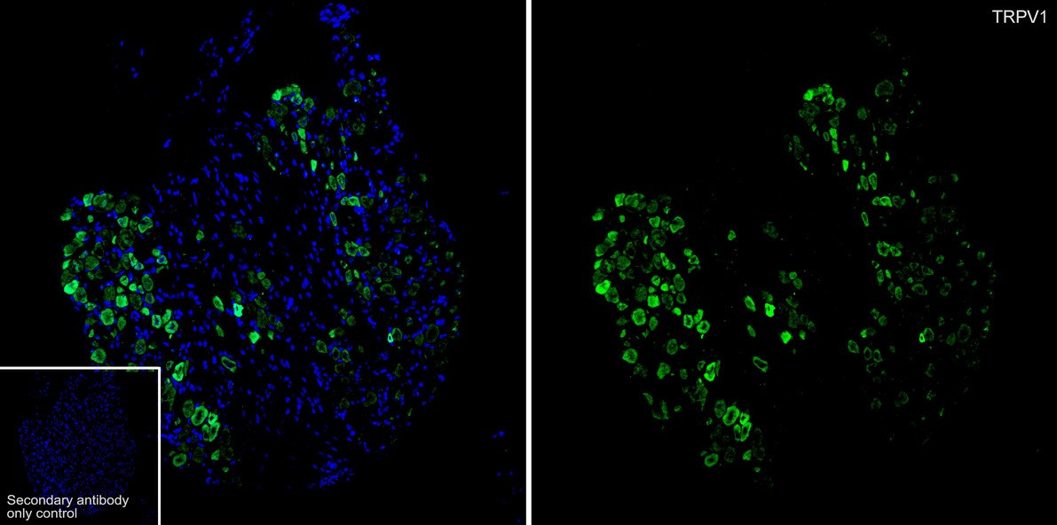
Application: IF-Tissue. Species: Mouse. Site: dorsal root ganglion. Sample: Paraffin-embedded section. Antibody concentration: 1/10,000.IF-P
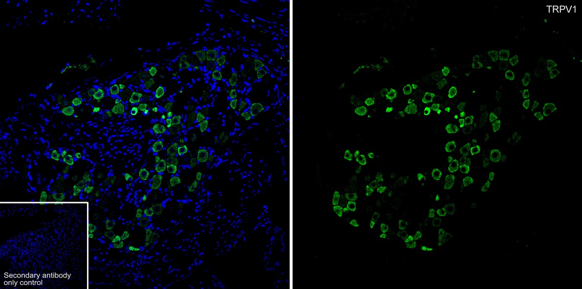
Application: IF-Tissue. Species: Rat. Site: dorsal root ganglion. Sample: Paraffin-embedded section. Antibody concentration: 1/15,000.| Product Name | TRPV1 Recombinant Rabbit Monoclonal Antibody |
|---|---|
| Antibody Type | Primary Antibodies |
| Immunogen | Recombinant protein within human TRPV1 aa 94-433. |
| Clonality | Monoclonal |
|---|---|
| Isotype | IgG |
| Host Species | Rabbit |
| Tested Applications | IF-FIF-PIHC |
| IHC:1:15000 IF-F:1:1000-1:5000 IF-P:1:10000-1:15000 |
|
| Species Reactivity | MouseRat |
| Concentration | 1mg/ml |
| Purification | Protein A |
| Gene Symbol | TRPV1 |
|---|---|
| Gene Synonyms | VR1 |
| Gene Full Name | transient receptor potential cation channel subfamily V member 1 |
| Gene Summary | Capsaicin, the main pungent ingredient in hot chili peppers, elicits a sensation of burning pain by selectively activating sensory neurons that convey information about noxious stimuli to the central nervous system. The protein encoded by this gene is a receptor for capsaicin and is a non-selective cation channel that is structurally related to members of the TRP family of ion channels. This receptor is also activated by increases in temperature in the noxious range, suggesting that it functions as a transducer of painful thermal stimuli in vivo. Four transcript variants encoding the same protein, but with different 5' UTR sequence, have been described for this gene. [provided by RefSeq, Jul 2008] |
| Alternative Names | Capsaicin receptor antibody DKFZp434K0220 antibody osm 9 like TRP channel 1 antibody Osm-9-like TRP channel 1 antibody OTRPC1 antibody Transient receptor potential cation channel subfamily V member 1 antibody TRPV 1 antibody Trpv1 antibody TRPV1_HUMAN antibody Vanilloid receptor 1 antibody Vanilloid receptor subtype 1 antibody VR 1 antibody VR1 antibody |
| Molecular Weight(MW) | 95 kDa |
| Cellular Localization | Postsynaptic cell membrane, Cell projection, dendritic spine membrane, Cell membrane. |
IHC
Immunohistochemical analysis of paraffin-embedded mouse dorsal root ganglion tissue with Rabbit anti-TRPV1 antibody at 1/15,000 dilution. The section was pre-treated using heat mediated antigen retrieval with Tris-EDTA buffer (pH 9.0) for 20 minutes. The tissues were blocked in 1% BSA for 20 minutes at room temperature, washed with ddH2O and PBS, and then probed with the primary antibody at 1/15,000 dilution for 1 hour at room temperature. The detection was performed using an HRP conjugated compact polymer system. DAB was used as the chromogen. Tissues were counterstained with hematoxylin and mounted with DPX.
IHC
Immunohistochemical analysis of paraffin-embedded rat dorsal root ganglion tissue with Rabbit anti-TRPV1 antibody at 1/15,000 dilution. The section was pre-treated using heat mediated antigen retrieval with Tris-EDTA buffer (pH 9.0) for 20 minutes. The tissues were blocked in 1% BSA for 20 minutes at room temperature, washed with ddH2O and PBS, and then probed with the primary antibody at 1/15,000 dilution for 1 hour at room temperature. The detection was performed using an HRP conjugated compact polymer system. DAB was used as the chromogen. Tissues were counterstained with hematoxylin and mounted with DPX.
IF-F
Application: IHC-Fr. Species: Mouse. Site: dorsal root ganglion. Sample: Frozen section. Antibody concentration: 1/1,000. Antigen retrieval: Not required.
IF-F
Application: IHC-Fr. Species: Rat. Site: dorsal root ganglion. Sample: Frozen section. Antibody concentration: 1/5,000. Antigen retrieval: Not required.
IF-P
Application: IF-Tissue. Species: Mouse. Site: dorsal root ganglion. Sample: Paraffin-embedded section. Antibody concentration: 1/10,000.
IF-P
Application: IF-Tissue. Species: Rat. Site: dorsal root ganglion. Sample: Paraffin-embedded section. Antibody concentration: 1/15,000.| Application Notes | IHC:1:15000 IF-F:1:1000-1:5000 IF-P:1:10000-1:15000 |
|---|
| Form | Liquid |
|---|---|
| Storage Instructions | Shipped at 4℃. Store at +4℃ short term (1-2 weeks). Store at -20℃ long term. |
| Storage Buffer | 1*PBS (pH7.4), 0.1% BSA, 40% Glycerol, 0.2% Proclean 950. |


